5.2 The Cellular Basis of Nutrient Absorption in Animals
Hannah Nelson and Christelle Sabatier
Learning Objectives
By the end of this section, you will be able to do the following:
- Describe the impact of surface area on nutrient absorption.
- Infer the type of transport needed for a molecule to cross the membrane based on a cell diagram.
- Infer the type of transport needed for a molecule to cross the membrane based on molecular structure.
Animals obtain their nutrition mostly from the consumption of other organisms. Depending on their diet, animals can be classified into the following categories: plant eaters (herbivores), meat eaters (carnivores), and those that eat both plants and animals (omnivores). Once consumed, nutrients and macromolecules present in food are not immediately accessible to the cells. There are a number of processes that modify food within the animal body in order to make the nutrients and organic molecules usable for cellular function. As animals evolve in form and function, their digestive systems have also evolved to accommodate their various dietary needs. Digestion involves more than just material—it is a complex system of breakdown and nutrient transfer that will fuel or fail the body.
Once the macromolecules in the food are converted into monomers, these monomers can be absorbed into the bloodstream so that they can be distributed to all cells in the body to support cell maintenance and growth.
The stomach is the major site for protein digestion in animals other than ruminants (e.g., cows and deer). Protein digestion is mediated by an enzyme called pepsin in the stomach chamber. Pepsin is a protease secreted by cells in the stomach where it breaks peptide bonds and cleaves proteins into smaller polypeptides and amino acids. Another cell type—parietal cells—secrete hydrogen and chloride ions, which combine in the lumen to form hydrochloric acid, the primary acidic component of the stomach juices. The highly acidic environment kills many microorganisms in the food and, combined with the action of the enzyme pepsin, results in the hydrolysis of protein in the food.
We can look at the parietal cell, which is responsible for producing the acid (HCl) in the stomach to infer the type of membrane transport responsible for ensuring that H+ and Cl– are in high concentrations in the stomach lumen (Figure 5.2.1).
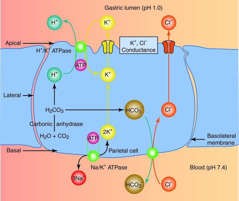
Inferring concentration of H+ in the parietal cell: The enzyme carbonic anhydrase converts one molecule of carbon dioxide and one molecule of water indirectly into a bicarbonate ion (HCO3–) and a hydrogen ion (H+). This leads to a high concentration of H+ in the cytoplasm of the parietal cell.
Primary active transport of H+ into the gastric lumen: In the stomach ion exchange is used to move H+ ions out the cells and into the lumen of the stomach. The H+/K+ ATPase uses the energy from the hydrolysis of ATP to push H+ out of the cell and pull K+ into the cell. This ensures that there is no net charge build up across the membrane and the highest concentration of H+ is found in the gastric lumen.
Secondary active transport of Cl– into the parietal cell: The bicarbonate ion (HCO3–), which builds up in the parietal cell cytoplasm, is then exchanged for a chloride ion (Cl–) on the basal side (away from the lumen) of the cell. The transport of HCO3– down its concentration gradient provides the energy to drive Cl– ions into the parietal cell cytoplasm. Cl– ions can then diffuse through a channel into the gastric lumen side of the cell.
When digesting protein and some fats, the stomach lining must be protected from getting digested by pepsin. The stomach lining protects itself by synthesizing the pepsin enzyme in the inactive form to protect chief cells and maintaining a thick mucus lining that protects the underlying tissue from digestive juices.
Small Intestine
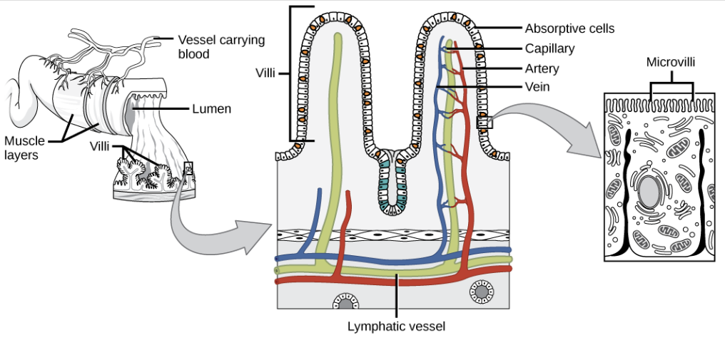
Chyme moves from the stomach to the small intestine. The small intestine is the organ where the digestion of protein, fats, and carbohydrates is completed. The small intestine is a long tube-like organ with a highly folded surface containing finger-like projections called the villi. The apical surface of each villus has many microscopic projections called microvilli. These structures, illustrated in Figure 5.2.2, are lined with epithelial cells on the luminal side (facing the interior of the small intestine) and allow for the nutrients to be absorbed from the digested food and absorbed into the bloodstream on the other side. The villi and microvilli, with their many folds, increase the surface area of the intestine and increase absorption efficiency of the nutrients. Absorbed nutrients in the blood are carried into the hepatic portal vein, which leads to the liver. There, the liver regulates the distribution of nutrients to the rest of the body and removes toxic substances, including drugs, alcohol, and some pathogens.
Practice Questions
Large Intestine
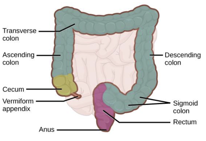
The large intestine, illustrated in Figure 5.2.3, reabsorbs the water from the undigested food material and processes the waste material. The human large intestine is much smaller in length compared to the small intestine but larger in diameter. The colon is home to many bacteria or “intestinal flora” sometimes referred to as “gut flora” that aid in the digestive processes. The main functions of the colon are to extract the water and mineral salts from undigested food, and to store waste material. Carnivorous mammals have a shorter large intestine compared to herbivorous mammals due to their diet (Figure 5.2.3).
Consider This: An Increasing Interest in Insect Protein
Insects are high in protein, fiber, and vitamins. Recently, American food producers have begun to fund more research into this more environmentally friendly nutritional source for snack foods. Tyson foods, an Arkansas based company, as well as several startups across the country are developing insect based high protein products. The United States is a bit late to the trade, but pursuing high level investments in the opportunity to lower environmental waste.
Abundance and high nutritional value have made invertebrates like crickets and silkworms relatively common in the diet of some parts of Africa and East Asia. For example, Thailand and Cambodia are known for their street food, a staple of which is the incorporation of crickets. Moreover, bombyx mori—silkworm larvae—are a delicacy in Korea, Japan, and China.
The commonality of silkworms and crickets in certain eastern foods aligns with their cultural significance. The constant chirping of crickets brings good luck in Chinese culture, and silkworms are symbols of diligence and a dynamic spirit.
Practice Questions
Glossary
lumen
the cavity of a tubular organ (e.g. gut lumen) or organelle (e.g. vesicle lumen)
villi
small, finger-like projections made up of epithelial cells and blood vessels that extend into the lumen of the small intestine
microvilli
microscopic membrane projections from epithelial cells lining the intestinal lumen
small intestine
the organ where the digestion of protein, fats, and carbohydrates is completed and absorption of their monomers takes place
large intestine
reabsorbs the water from the undigested food material and processes the waste material
Figure Descriptions
Figure 5.2.1. A single blue parietal cell sits between a pink region at the top labeled “Gastric lumen (pH 1.0)” and a peach region at the bottom labeled “Blood (pH 7.4).” Orientation labels read Apical at the top, Basal at the bottom, Lateral on the side, and Basolateral membrane along the right edge. Inside the cell, the words “Carbonic anhydrase” appear beside the reaction “H₂O + CO₂ → H₂CO₃,” with arrows showing H⁺ (green) rising upward and HCO₃⁻ (brown) moving toward the basolateral side. On the apical surface to the left, a purple complex labeled “H⁺/K⁺ ATPase” (with a small ATP circle) shows H⁺ moving out to the lumen while K⁺ (yellow) moves in; a nearby opening labeled “K⁺, Cl⁻ conductance” returns K⁺ to the lumen. On the apical surface to the right, a red pathway marked Cl⁻ carries chloride into the gastric lumen. Along the basolateral membrane, a green transport shape shows HCO₃⁻ moving to the blood and Cl⁻ (red) moving into the cell. At the basal region, a complex labeled “Na⁺/K⁺ ATPase” (with ATP) displays “3Na⁺” leaving the cell and “2K⁺” entering. The colored spheres—green H⁺, yellow K⁺, red Cl⁻, and brown HCO₃⁻—and the arrows together depict the production of hydrochloric acid at the cell’s apical surface. [Return to Figure 5.2.1]
Figure 5.2.2. The figure moves from wide to close view to show how surface area increases for absorption. At left, a cutaway of the small intestine labels muscle layers, the interior lumen, and tuft-like villi projecting from the lining; a small vessel is marked “vessel carrying blood.” The center panel enlarges a section of lining into two tall villi with a thin epithelium of absorptive cells; inside each villus run a red artery, blue vein, and a thin capillary network, while a pale green lymphatic vessel (lacteal) lies in the core and along the base. The top surface facing the lumen is densely fringed, and a right-hand inset zooms further into one absorptive cell to show a brush border of tiny microvilli. Together the panels depict villi and microvilli as folds that greatly expand the intestinal surface for nutrient uptake into blood and lymph. [Return to Figure 5.2.2]
Figure 5.2.3. A simplified digestive-tract diagram highlights the large intestine in teal looping around a faint pink small intestine. Starting at the lower right side is the bulbous cecum (shown in yellow) with a thin vermiform appendix attached; from there the tube rises as the ascending colon, turns across the top as the transverse colon, descends on the opposite side as the descending colon, and curves into a purple sigmoid colon that continues as the rectum and ends at the anus. Black leader lines point to each part, illustrating the pathway where water is reabsorbed and waste is stored before elimination. [Return to Figure 5.2.3]
Licenses and Attributions
“5.2 Nutrient Absorption in Animals” is adapted from “34.1 Digestive Systems” by Mary Ann Clark, Matthew Douglas, and Jung Choi for OpenStax Biology 2e under CC-BY 4.0. “5.2 Nutrient Absorption in Animals” is licensed under CC-BY-NC 4.0.
Media Attributions
- 1A.B.4 Parietal Cell © Cenveo is licensed under a CC BY-NC (Attribution NonCommercial) license
- 1A.B.complex intestinal villi
- 1A.B.large intestine absorption
the cavity of a tubular organ (e.g. gut lumen) or organelle (e.g. vesicle lumen)
A mix of partially digested food and gastric juices produced in the stomach.
the organ where the digestion of protein, fats, and carbohydrates is completed and absorption of their monomers takes place
small, finger-like projections made up of epithelial cells and blood vessels that extend into the lumen of the small intestine
microscopic membrane projections from epithelial cells lining the intestinal lumen
reabsorbs the water from the undigested food material and processes the waste material
