10.1 Cellular Respiration Overview
Christelle Sabatier
Learning Objectives
By the end of this section, you will be able to do the following:
- Identify the major products of each step of aerobic cellular respiration.
- Compare and contrast the energy transformations that take place during aerobic cellular respiration.
- Identify the specific locations where each step of aerobic cellular respiration takes place.
C6H12O6 + 6O2 → 6CO2 + 6H2O + energy
The overall reaction for cellular respiration should look familiar. In Chapter 7, we explored in detail the energy transformations that transformed energy from sunlight into the energy captured in the C-C bonds of glucose. Glucose and other forms of carbohydrates allow this energy to be stored and transported within and between organisms through stable molecules. Cellular respiration is the process by which that energy is extracted and used to drive work in cells. All cells, prokaryotic and eukaryotic, undergo cellular respiration.
Oxidation-Reduction Reactions
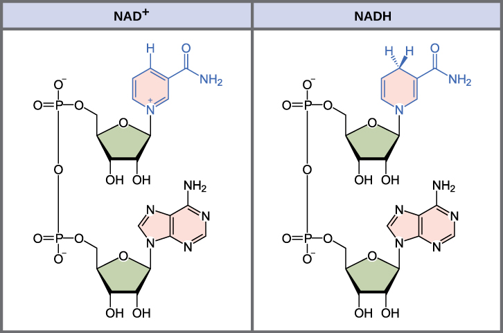
To transfer the energy from C-C bonds to the terminal phosphate bond of ATP, electrons must be sequentially extracted from the glucose molecule. This is done through a sequence of oxidation-reduction or redox reactions where the oxidized carbon donates electrons to electron carriers that become reduced in the process. The primary electron carrier in cellular respiration is nicotinamide adenine dinucleotide (NAD) (Figure 10.1.1), which is derived from vitamin B3, niacin. NAD+ is the oxidized form of the moleculeNAD+; NADH is the reduced form of the molecule after it has accepted two electrons and a proton (which together are the equivalent of a hydrogen atom with an extra electron). Note that if a compound has an “H” on it, it is generally reduced (e.g., NADH is the reduced form of NAD).
Mitochondria and Cellular Respiration
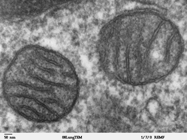
In prokaryotes, all major steps of cellular respiration take place in the cytoplasm or the plasma membrane. However, in eukaryotes, some of the steps take place in a dedicated organelle, the mitochondrion. Mitochondria are oval-shaped, double membrane organelles (Figure 10.1.2) that have their own ribosomes and DNA. Each membrane is a phospholipid bilayer embedded with proteins. The inner mitochondrial membrane has many folds to maximize surface area for some of the processes of cellular respiration. We call the area surrounded by the folds the mitochondrial matrix, where additional steps in cellular respiration take place.
Steps of Cellular Respiration
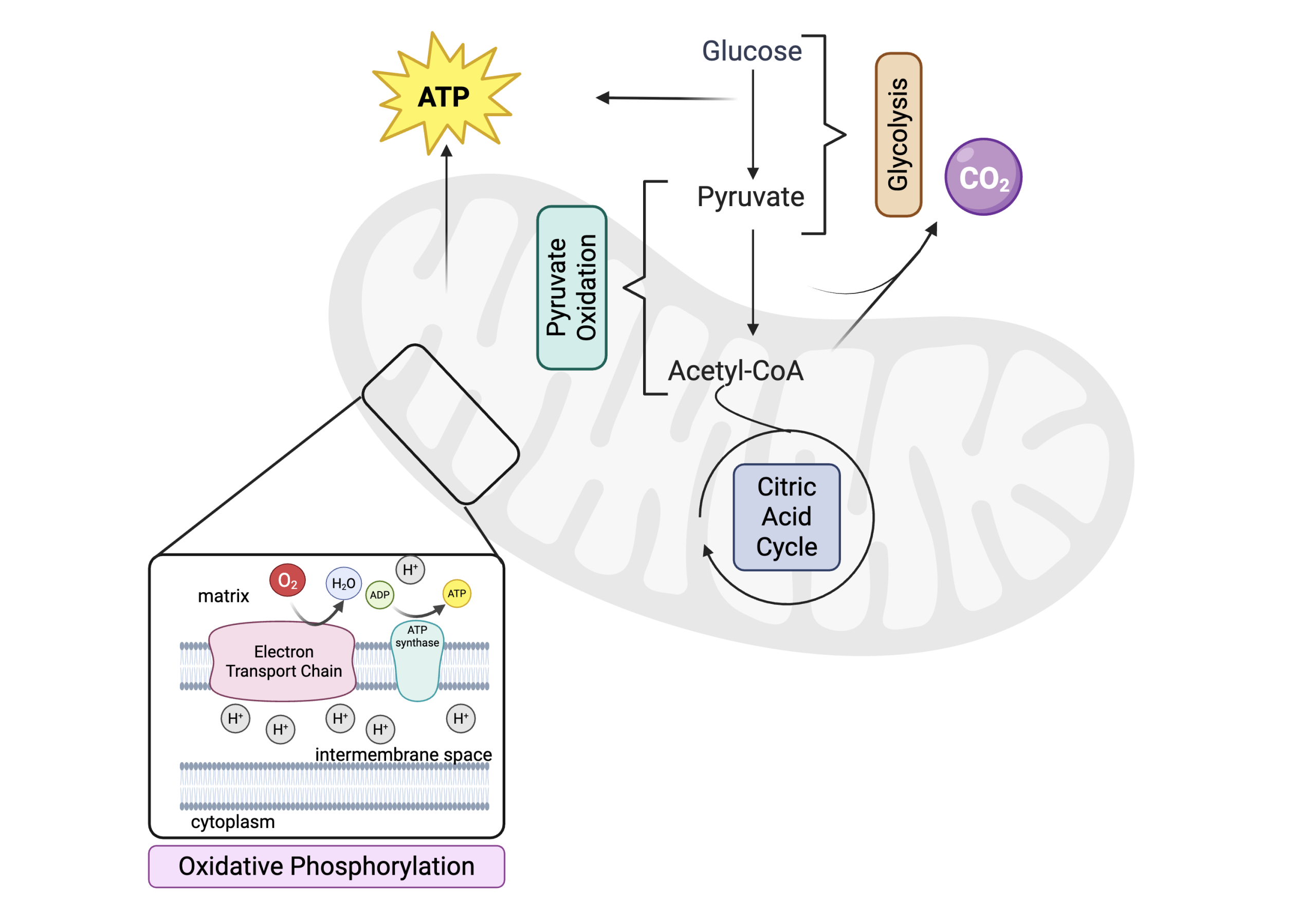
Cellular respiration is made up of 4 major steps (Figure 10.1.3). Below is an overview of these steps that are described in more details in the subsequent sections:
- Glycolysis where the 6-C glucose is oxidized into two molecules of 3-C pyruvate. This takes place in the cytoplasm and produces NADH and ATP (see 10.2 Glycolysis).
- Pyruvate oxidation produces NADH, the first molecules of CO2, and a 2-C acetate molecule, which is coupled to coenzyme A to form acetyl-coA. This step takes place between the cytoplasm where pyruvate is produced and the mitochondrial matrix where acetyl-coA is produced. (see 10.3 Pyruvate Oxidation and the Citric Acid Cycle)
- Acetyl-coA then enters the citric acid cycle, a multi-step process where the last carbons from the original glucose molecule are oxidized into CO2. Many reduced electron carriers are produced in this process in the mitochondrial matrix. (see 10.3 Pyruvate Oxidation and the Citric Acid Cycle)
- The electron carriers produced in the previous steps donate their electrons to the electron transport chain located on the inner membrane of the mitochondrion. These high energy electrons are passed between molecular complexes embedded in the membrane until they are ultimately donated to O2, which is reduced to H2O. The electron movement through the chain provides energy to pump protons (H+) from the matrix into the intermembrane space. (see 10.4 Oxidative Phosphorylation)
- The electron gradient across the inner mitochondrial membrane drives the activity of ATP synthase, which catalyzes the formation of ATP from ADP and phosphate as protons diffuse back into the mitochondrial matrix. This is where the majority of ATP is produced in cellular respiration. (see 10.4 Oxidative Phosphorylation)
Practice Questions
Glossary
cellular respiration
metabolic pathway that transfer the energy in the C-C bonds of glucose to the molecule ATP
oxidation-reduction/redox reaction
oxidized carbon donates electrons to electron carriers that become reduced in the process
NAD
nicotinamide adenine dinucleotide, derived from niacin
NADH+
oxidized form of NAD
NADH
reduced form of NAD
mitochondrial matrix
internal spaces of mitochondrion enclosed by the inner membrane
Figure Descriptions
Figure 10.1.1. The image is a diagram comparing the chemical structures of NAD⁺ and NADH placed side by side in two separate panels. On the left, the structure of NAD⁺ is shown, containing two ribose sugar molecules (displayed in green), two nucleotides with phosphate groups, and a nicotinamide ring indicated in pink and blue. The nicotinamide in NAD⁺ has a positive charge and is represented with a plus sign. On the right, the structure of NADH is nearly identical but with a slight difference in the nicotinamide ring, where a hydrogen atom is added, and the positive charge is absent. [Return to Figure 10.1.1]
Figure 10.1.2. The image is a grayscale transmission electron microscope (TEM) micrograph showing a close-up view of two mitochondria. Each mitochondrion appears as an oval shape with a defined outer boundary. Inside, distinct, wave-like cristae are visible, giving the internal structure a layered appearance. The background consists of a textured, grainy pattern typical of electron micrographs. The image scale is indicated at the bottom, showing 50 nm. [Return to Figure 10.1.2]
Figure 10.1.3. The image is a flowchart illustrating cellular respiration within a stylized mitochondrion. At the top, a yellow starburst labeled “ATP” indicates the production of ATP. The process begins with Glucose, which converts to Pyruvate through Glycolysis. Pyruvate is further converted to Acetyl-CoA through pyruvate oxidation. Then Acetyl-coA enters the Citric Acid Cycle in the mitochondrial matrix. A lavender circle labeled “CO₂” shows carbon dioxide release from pyruvate oxidation and the citric acid cycle. Oxidative phosphorylation is shown in the inner mitochondrial membrane, enlarged in a cutout. The cutout displays the Electron Transport Chain (ETC), where O₂ converts to H₂O, and ATP synthase produces ATP from ADP and phosphate in the matrix. The ETC and ATP synthase are both located in the inner mitochondrial membrane and contribute to or rely on a proton (H+) gradient where there are more protons in the intermembrane space than in the matrix. [Return to Figure 10.1.3]
Licenses and Attributions
“10.1 Cellular Respiration Overview” is adapted from “7.1 Energy in Living Systems” by Mary Ann Clark, Matthew Douglas, and Jung Choi for OpenStax Biology 2e under CC-BY 4.0. “10.1 Cellular Respiration Overview” is licensed under CC-BY-NC 4.0.
Media Attributions
- 1A.10.1NADH © OpenStax Biology 2e is licensed under a CC BY (Attribution) license
- Mitochondria,_mammalian_lung_-_TEM © Louise Howard is licensed under a Public Domain license
- 1A.10.1Cellular Respiration © BioRender adapted by Christelle Sabatier is licensed under a Public Domain license
metabolic pathway that transfer the energy in the C-C bonds of glucose to the molecule ATP
cell that lacks a distinct nucleus and other organelles due to the absence of internal membranes.
any cell that possesses a clearly defined nucleus.
high energy molecule used as currency for cellular processes
oxidized carbon donates electrons to electron carriers that become reduced in the process
nicotinamide adenine dinucleotide, derived from niacin
oxidized form of NAD
reduced form of NAD
internal spaces of mitochondrion enclosed by the inner membrane
