21.4 Signaling Insulin Release
Learning Objectives
By the end of this section, you will be able to do the following:
- Apply the principles of ion channel-linked receptors signaling to how insulin is releases from pancreatic beta cells.
Ion channel-linked receptors
Ion channel-linked receptors bind a ligand and open a channel through the membrane that allows specific ions to pass through. To form a channel, this type of cell-surface receptor has an extensive membrane-spanning region. In order to interact with the phospholipid fatty acid tails that form the center of the plasma membrane, many of the amino acids in the membrane-spanning region are hydrophobic in nature. Conversely, the amino acids that line the inside of the channel are hydrophilic to allow for the passage of water or ions. These channels can bind ligands on either the extracellular side or on the cytoplasmic side of the membrane. In either case, ligand binding results in a conformational change in the protein’s structure that regulates the flow of ions such as sodium, calcium, or potassium to pass through. This form of signaling can have a very quick impact on the membrane potential across the membrane, which may result in quick responses. This is often seen in neuronal signaling pathways and, as discussed below, is a strategy used to trigger the release of insulin from pancreatic beta cells in response to the increase in blood glucose levels past the set point.
Glucose-dependent Release of Insulin
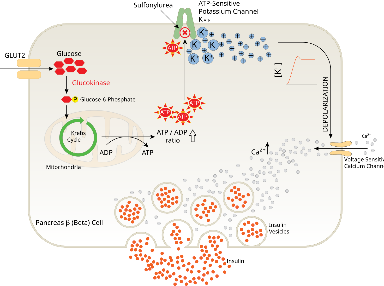
As a peptide hormone, insulin is produced through the secretory pathway in pancreatic beta cells. While blood glucose levels are at or below their set point, insulin is held in internal vesicles in the beta cells (Figure 1). They can be released through the process of exocytosis (you can review what that process looks like in Section 14.7 The endomembrane system). We will explore the steps that connect the stimulus (blood glucose concentration increase) to the response (insulin release).
Glucose concentration sensing
Pancreatic beta cells express the glucose transporter (GLUT2). Glucose quickly equilibrates between its concentration in the blood stream and its concentration in the cytoplasm of pancreatic beta cells by flowing through GLUT2. Inside the beta cells, glucose is phosphorylated by glucokinase, the first enzyme in glycolysis and cellular respiration. Ultimately, energy contained in the C-C bonds of glucose is extracted and used to drive the formation of ATP from ADP and inorganic phosphate (Pi).
When glucose levels flowing into the cell increase, the ATP/ADP ratio increases as more ATP is produced and levels of ADP decrease.
Change in ion balance across the beta cell membrane
An ATP-sensitive potassium channel regulates the membrane potential across the membrane of the beta cell. This can be measured in millivolts (mV) and can be thought of like a battery, a measure of the separation of positive and negative charges across the membrane. When glucose levels outside the cell are low, this channel is open, allowing K+ ions to flow out of the cell and keeping the inside of the cell more negatively charged. As glucose levels rise so do ATP levels inside the cell. ATP leads to the closing of the ATP-sensitive potassium channel, blocking the path for K+ ions to flow out of the cell. This results in more positive charge remaining in the cell also known as a depolarization of the plasma membrane.
Insulin exocytosis
This depolarization triggers the opening of a voltage-sensitive Ca2+ channel, which allows Ca2+ ions to flow from the outside of the cell into the cytoplasm, down their electrochemical gradient. Ca2+, which can also serve as a second messenger, binds to a protein on the insulin filled vesicles and triggers their fusion with the plasma membrane. The fusion of the vesicle membrane and the plasma membrane is the process of exocytosis that allows insulin hormones to be released out of the beta cells and into the extracellular fluid. Very quickly, the insulin will diffuse into nearby blood vessels where they will flow around the body and have the potential to trigger responses from cells expressing the insulin receptor.

Figure 1. The mechanism of glucose-dependent insulin release in pancreatic Beta cells.
Figure Descriptions
Figure 1. The image is a diagram illustrating the process of insulin release from a pancreatic beta cell. In the top left, glucose molecules are shown entering the cell where they are converted into glucose-6-phosphate by glucokinase. This glucose-6-phosphate enters the Krebs Cycle within the mitochondria, producing ATP. The diagram indicates an increase in the ATP/ADP ratio. ATP molecules interact with the potassium ion (K+) channels on the cell membrane, leading to channel closure and depolarization of the membrane. This is indicated by a graph showing a rise in [K+]. The depolarization allows calcium ions (Ca2+) to enter the cell, leading to the release of insulin from vesicles situated at the bottom of the diagram. The vesicles are depicted releasing their contents outside the cell.
Media Attributions
- Glucose_Insulin_Release_Pancreas.svg © aydintay is licensed under a CC BY (Attribution) license
