4.1 Cell Types and Compartments
Christelle Sabatier
Learning Objectives
By the end of this section, you will be able to do the following:
- Describe the role of cells in organisms.
- Compare and contrast prokaryotic and eukaryotic cells.
- Describe the relative sizes of different cells.
- Explain why cells must be small.
A cell is the smallest unit of a living thing. Organisms are either comprised of one cell (like bacteria) or many cells (like a human). Thus, cells are the basic building blocks of all organisms.
Several cells of one kind that interconnect with each other and perform a shared function form tissues. These tissues combine to form an organ (your stomach, heart, or brain), and several organs comprise an organ system (such as the digestive system, circulatory system, or nervous system). Several systems that function together form an organism (like a human being). Here, we will examine the structure and function of cells.
There are many types of cells, which scientists group into one of two broad categories: prokaryotic and eukaryotic. For example, we classify both animal and plant cells as eukaryotic cells; whereas, we classify bacterial cells as prokaryotic.
All cells share four common components: 1) a plasma membrane, an outer covering that separates the cell’s interior from its surrounding environment; 2) cytoplasm, consisting of a jelly-like cytosol within the cell in which there are other cellular components; 3) DNA, the cell’s genetic material; and 4) ribosomes, which synthesize proteins.
Prokaryotic Cells
A prokaryote is a simple, mostly single-celled (unicellular) organism that lacks a nucleus, or any other membrane-bound organelle. We will shortly come to see that this is significantly different in eukaryotes. Prokaryotic DNA is in the cell’s central part: the nucleoid (Figure 4.1.1).
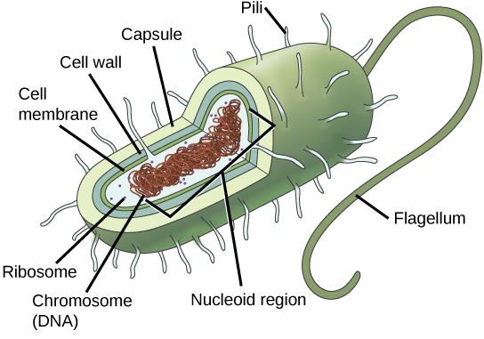
In addition to a plasma membrane, most prokaryotic cells have a peptidoglycan cell wall and many have a polysaccharide capsule (Figure 4.1.1). The cell wall acts as an extra layer of protection, helps the cell maintain its shape, and prevents dehydration. The capsule enables the cell to attach to surfaces in its environment. Some prokaryotes have flagella, pili, or fimbriae. Flagella are used for locomotion. Pili exchange genetic material during conjugation, the process by which one bacterium transfers genetic material to another through direct contact. Bacteria use fimbriae to attach to a host cell.
Cell Size
At 0.1 to 5.0 micrometer (μm) in diameter, prokaryotic cells are significantly smaller than eukaryotic cells, which have diameters ranging from 10 to 100 μm. The prokaryotes’ small size allows ions and organic molecules that enter them to quickly diffuse to other parts of the cell. Similarly, any wastes produced within a prokaryotic cell can quickly diffuse. This is not the case in eukaryotic cells, which have developed different structural adaptations to enhance intracellular transport.
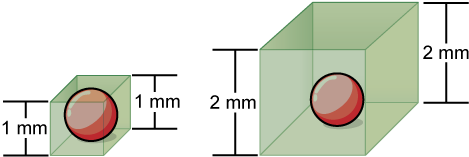
Small size, in general, is necessary for all cells, whether prokaryotic or eukaryotic. Let’s examine why that is so. First, we’ll consider the area and volume of a typical cell. Not all cells are spherical in shape, but most tend to approximate a sphere. You may remember from your high school geometry course that the formula for the surface area of a sphere is 4πr2, while the formula for its volume is 4πr3/3. Thus, as the radius of a cell increases, its surface area increases as the square of its radius, but its volume increases as the cube of its radius (much more rapidly). Therefore, as a cell increases in size, its surface area-to-volume ratio decreases. This same principle would apply if the cell had a cube shape (Figure 4.1.2). If the cell grows too large, the plasma membrane will not have sufficient surface area to support the rate of diffusion required for the increased volume. In other words, as a cell grows, it becomes less efficient. One way to become more efficient is to divide. Other ways are to increase surface area by foldings of the cell membrane, become flat or thin and elongated, or develop organelles that perform specific tasks. These adaptations lead to developing more sophisticated cells, which we call eukaryotic cells.
Eukaryotic cells
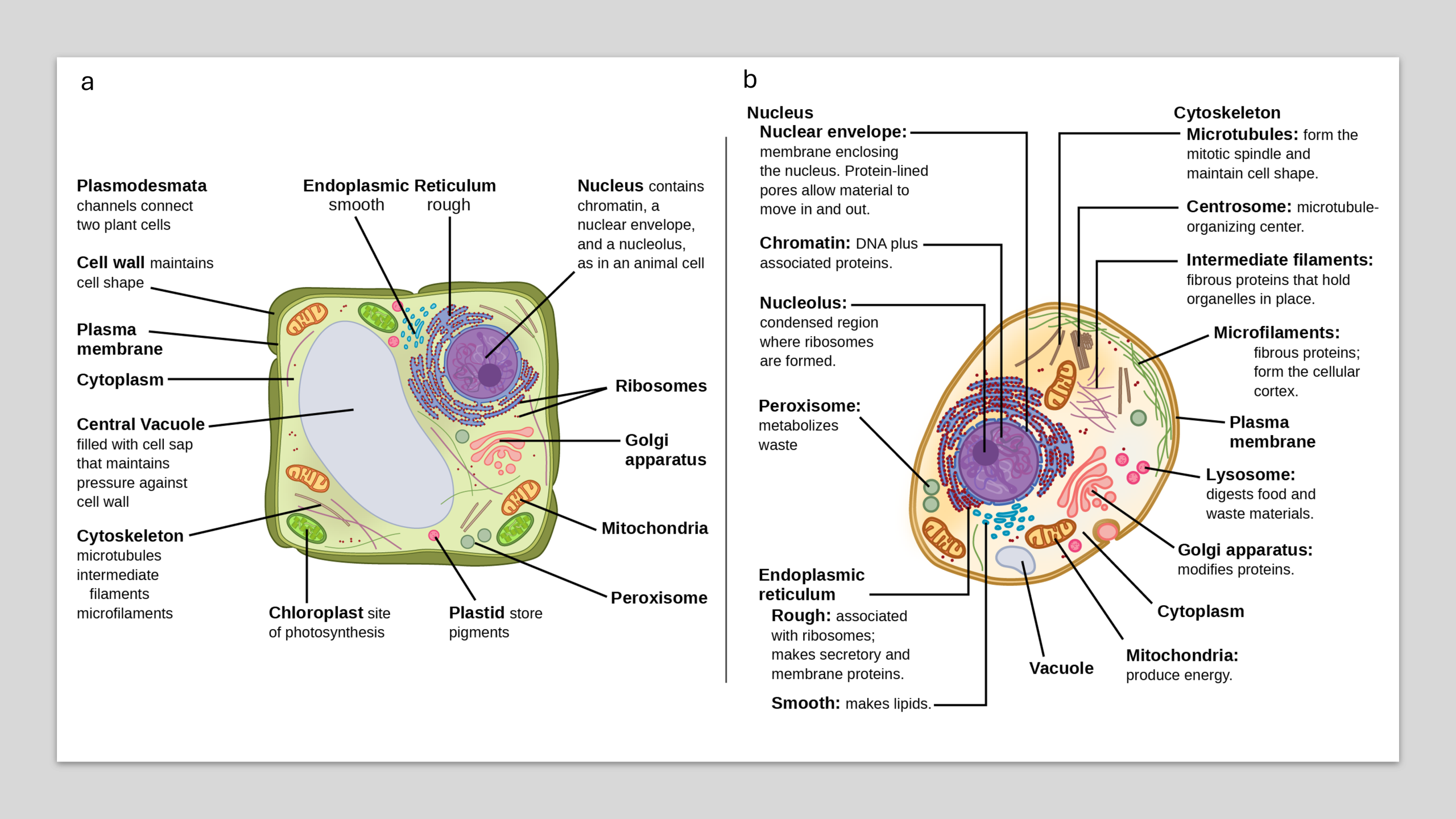
Unlike prokaryotic cells, eukaryotic cells have: 1) a membrane-bound nucleus; 2) numerous membrane-bound organelles such as the endoplasmic reticulum, Golgi apparatus, chloroplasts, mitochondria, and others; and 3) several, rod-shaped chromosomes. Because a membrane surrounds eukaryotic cell’s nucleus, it has a “true nucleus.” The word “organelle” means “little organ,” and, as we already mentioned, organelles have specialized cellular functions, just as your body’s organs have specialized functions.
At this point, it should be clear to you that eukaryotic cells have a more complex structure than prokaryotic cells. Organelles allow different functions to be compartmentalized in different areas of the cell. And not every eukaryotic cell has the same complement of organelles depending on its function. Even within the same organism, different cells will leverage different organells. For example, in plants chloroplasts are primarily found in the parts of the plant above ground that conduct photosynthesis while plastids, which store pigments and starch, are found in below ground structures. You will learn a lot more details about the individual organelles found in eukaryotes as you proceed through the introductory biology courses. For now, let’s first examine two important components of the cell: the plasma membrane and the cytoplasm.
The Cytoplasm
The cytoplasm is the cell’s entire region between the plasma membrane and the nuclear envelope (a structure we will discuss shortly). It is comprised of organelles suspended in the gel-like cytosol, the cytoskeleton, and various chemicals (Figure 4.1.3). Even though the cytoplasm consists of 70% to 80% water, it has a semisolid consistency, which comes from the proteins within it. However, proteins are not the only organic molecules in the cytoplasm. Glucose and other simple sugars, polysaccharides, amino acids, nucleic acids, fatty acids, and derivatives of glycerol are also there. Ions of sodium, potassium, calcium, and many other elements also dissolve in the cytoplasm. Many metabolic reactions, including protein synthesis, take place in the cytoplasm.
The Plasma Membrane
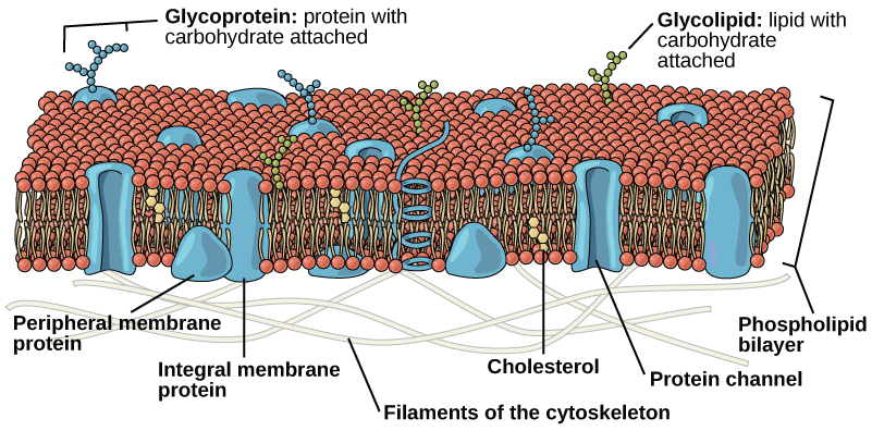
Like prokaryotes, eukaryotic cells have a plasma membrane (Figure 4.1.4), a phospholipid bilayer with embedded proteins that separates the internal contents of the cell from its surrounding environment. A phospholipid is a lipid molecule with two fatty acid chains and a phosphate-containing group. The plasma membrane controls the passage of organic molecules, ions, water, and oxygen into and out of the cell. Wastes (such as carbon dioxide and ammonia) also leave the cell by passing through the plasma membrane.
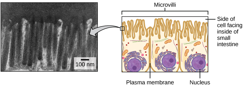
The plasma membranes of cells that specialize in absorption fold into fingerlike projections that we call microvilli (singular = microvillus); (Figure 4.1.5). Such cells typically line the small intestine, the organ that absorbs nutrients from digested food. This is an excellent example of form following function. People with celiac disease have an immune response to gluten, which is a protein in wheat, barley, and rye. The immune response damages microvilli, and thus, afflicted individuals cannot absorb nutrients. This leads to malnutrition, cramping, and diarrhea. Patients suffering from celiac disease must follow a gluten-free diet.
In addition, many of the organelles in eukaryotic cells are surrounded by a phospholipid bilayer. This allows very different environments to exist inside each organelle compared to the cytoplasm. Each environment is maximally suited to the specific function of that organelle. For example, lysosomes in animal cells and vacuoles in plant cells are the site where many macromolecules are recycled into their constituent monomers. Proteins are digested into individual amino acids that can then be recycled as new proteins are synthesized in the cytoplasm. The enzymes that are responsible for catalyzing these reactions function at low pH, so the interior of lysosomes and vacuoles are often much more acidic than the cytoplasm. This allows these processes to take place efficiently without interfering with other functions of the cells that might be disrupted by an acidic environment.
Overview of Key Eukaryotic Organelles
Practice Questions
Glossary
cells
smallest fundamental unit of structure and function in living organisms.
cytoplasm
semifluid substance of a cell that is external to the nuclear membrane and internal to the cellular membrane
eukaryotic cell
any cell that possesses a clearly defined nucleus.
organism
individual living entity
organelle
aggregates of macromolecules surrounded by membranes
plasma membrane
phospholipid bilayer that surrounds every living cell, delimiting the cell from the environment around it.
prokaryotic cell
cell that lacks a distinct nucleus and other organelles due to the absence of internal membranes.
Figure Descriptions
Figure 4.1.1. The image is a labeled diagram of a bacterium, illustrating its internal and external structures. The bacterium is depicted in an oblong, oval shape with a green outer layer representing the capsule. Several hair-like projections, called pili, emerge from the surface. Inside, the layers consist of a cell wall, cell membrane, and an inner area highlighted as the nucleoid region with coiled brown structures labeled as the chromosome (DNA). Scattered within the cell are smaller round structures labeled as ribosomes. Extending from one end of the bacterium is a long, whip-like structure labeled as the flagellum. [Return to Figure 4.1.1]
Figure 4.1.2. The image shows two transparent green cubes, each containing a red sphere inside. The cube on the left is smaller (1 mm on each side), and the sphere fits snugly inside it. The cube on the right is larger (2 mm on each side), with more space around the sphere, making it appear more spacious. [Return to Figure 4.1.2]
Figure 4.1.3. The image contains two labeled illustrations separated by a thin vertical line. Panel (a) on the left depicts a typical eukaryotic plant cell: a rectangular green outline represents the rigid cell wall, inside which a thinner plasma membrane encloses the pale-green cytoplasm. Prominent features include a large central vacuole filling most of the interior, green chloroplasts and other pigment-containing plastids, and numerous small peroxisomes; thin black arrows point to these structures and to plasmodesmata (channels through the wall), the smooth and rough endoplasmic reticulum, Golgi apparatus, ribosomes, mitochondria, and the purple nucleus with its envelope and nucleolus. Panel (b) on the right shows a roundish animal cell lacking a cell wall. Arrows label the nucleus (envelope, chromatin, nucleolus), an extensive rough and smooth ER, a stack-like Golgi, spherical lysosomes, a pair of centrosomes, and small vacuoles. The cytoskeleton is detailed: microtubules, intermediate filaments, and microfilaments radiate through the orange-rimmed cytoplasm, while multiple mitochondria and peroxisomes are scattered inside. The comparison highlights plant-specific structures—cell wall, chloroplasts, plastids, and a large central vacuole—and animal-specific ones such as lysosomes and centrosomes, illustrating shared eukaryotic organelles alongside kingdom-specific differences. [Return to Figure 4.1.3]
Figure 4.1.4. The image illustrates the structure of a cell membrane. It is a cross-sectional diagram showing a phospholipid bilayer, depicted by two rows of orange spheres with tails, representing lipids. Interspersed among the lipids are various components: blue, round-shaped proteins (integral and peripheral membrane proteins) embedded within or attached to the bilayer. Glycoproteins and glycolipids are shown with green carbohydrate chains extending outward. Cholesterol is depicted as small yellow molecules within the bilayer, influencing fluidity. The image also highlights protein channels extending across the membrane and filaments of the cytoskeleton below the bilayer. [Return to Figure 4.1.4]
Figure 4.1.5. The image consists of two main parts: an electron microscope photograph on the left and a detailed diagram on the right. The photograph shows a close-up view of the microvilli, depicted as dense, dark, vertical structures lined up closely, situated on the surface of a cell. The scale at the bottom right of the photograph indicates a measurement of 100 nanometers. The diagram on the right provides a colorful cross-sectional view of intestinal cells with their microvilli. It outlines three cells side by side, each featuring an outer layer of microvilli on the side facing the intestine. The microvilli are drawn as finger-like projections. Underneath the microvilli, the diagram shows components such as the plasma membrane, nucleus, as well as other organelles like mitochondria. Labels and arrows indicate the microvilli, plasma membrane, and describe that this is the side of the cell facing the inside of the small intestine. [Return to Figure 4.1.5]
Licenses and Attributions
“4.1 Cell Types and Compartments” is adapted by Christelle Sabatier from “4.2 Prokaryotic Cells” and “4.3 Eukaryotic Cells” by Mary Ann Clark, Matthew Douglas, and Jung Choi for OpenStax Biology 2e under CC-BY 4.0. “4.1 Cell Types and Compartments” is licensed under CC-BY-NC 4.0.
Media Attributions
- 1A.B.5 Prokaryotic cell © OpenStax Biology 2e is licensed under a CC BY (Attribution) license
- 1A.B.5 Surface Area to Volume ratio © OpenStax 2e is licensed under a CC BY (Attribution) license
- 1A.B-5-Comparing-eukaryotic-plant-and-animal-cells
- 1A.B.5 Plasma Membrane © OpenStax Biology 2e is licensed under a CC BY (Attribution) license
- 1A.B.5 Microvilli © Louisa Howard adapted by OpenStax Biology 2e is licensed under a CC BY (Attribution) license
phospholipid bilayer that surrounds every living cell, delimiting the cell from the environment around it.
semifluid substance of a cell that is external to the nuclear membrane and internal to the cellular membrane
cell that lacks a distinct nucleus and other organelles due to the absence of internal membranes.
any cell that possesses a clearly defined nucleus.
aggregate of macromolecules surrounded by membranes
a protein's monomer; has a central carbon or alpha carbon to which an amino group, a carboxyl group, a hydrogen, and an R group or side chain is attached; the R group is different for all 20 common amino acids
