"

14.1 Overview of the Central Dogma of Information Flow

Elizabeth Dahlhoff and Melissa Hardy

Learning Objectives

By the end of this section, you will be able to do the following:

  • Define a gene.
  • Describe the central dogma of information flow in biological systems.
  • Understand the difference between transcription and translation.

Genes are at the center of biological information flow

Illustration of DNA unwinding from chromosomes inside a cell (left) and a 3D protein structure of a membrane-spanning G-protein (right), labeled with helices. (linked Image Description available)

Figure 14.1. Genes, which are carried on chromosomes (left), are linearly organized instructions for making the RNA and protein molecules that are necessary for all of processes of life. A membrane-spanning G-protein (right) is just one example of the different molecular structures that are encoded by genes. (Image credits: “chromosome: Amoeba Sisters; “G protein”- Wikimedia Creative Commons). [Image Description]

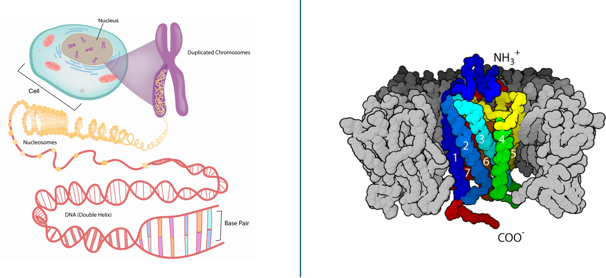
Since Gregor Mendel’s studies of peas over 100 years ago, the definition of a gene has progressed from an abstract unit of heredity to a tangible molecular entity capable of replication, expression, and mutation. Genes are composed of DNA and are linearly arranged on chromosomes. Genes specify sequences of amino acids that are the building blocks of proteins. In turn, proteins are responsible for orchestrating nearly every function of the cell. Both genes and the proteins they encode are absolutely essential to life as we know it.

The Central Dogma: DNA Encodes RNA; RNA Encodes Protein

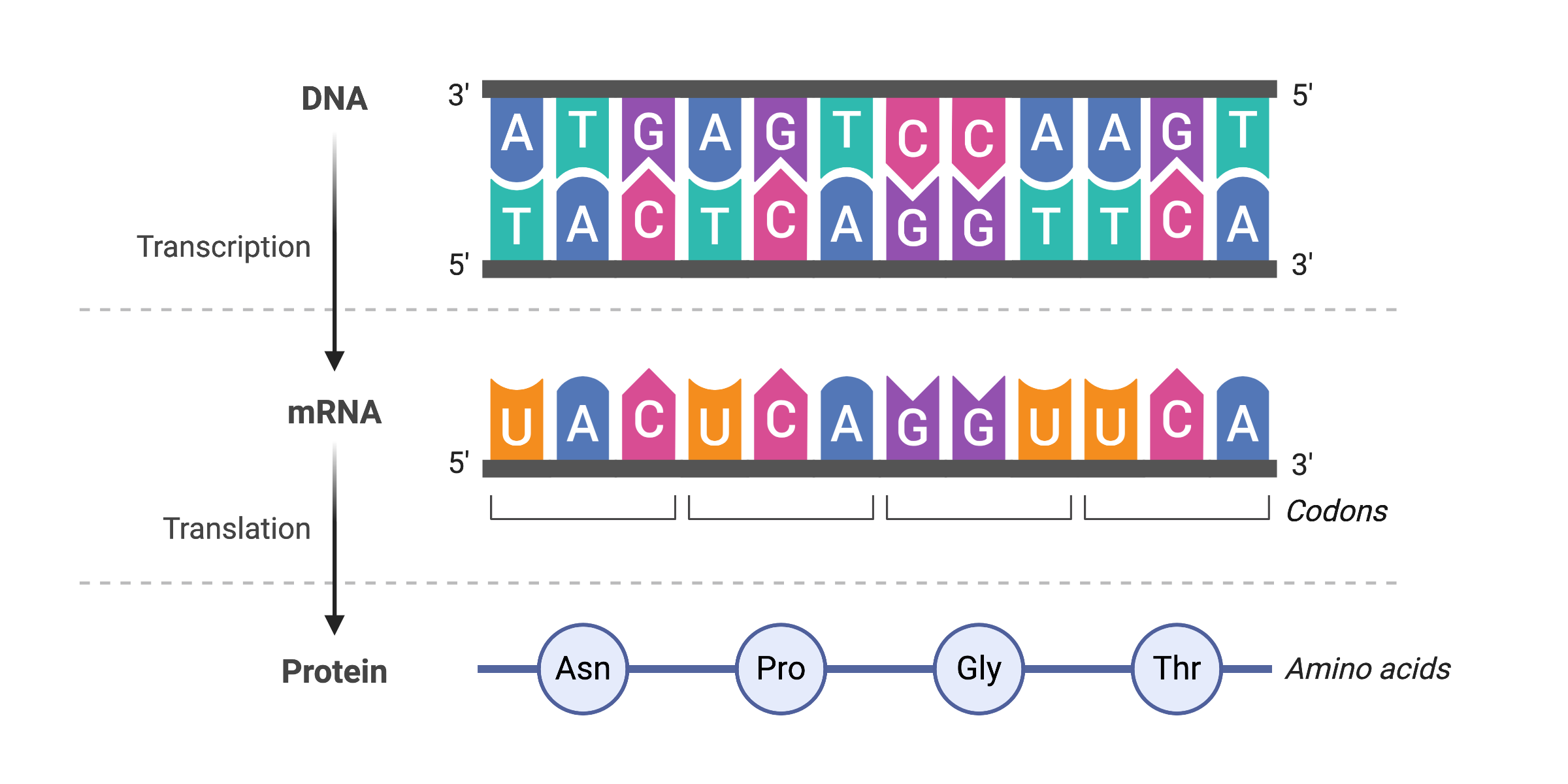
The flow of genetic information in cells from DNA to messenger RNA (mRNA) to protein is described by the central dogma, which states that genes specify the sequence of mRNAs, which in turn specify the sequence of amino acids (polypeptide) making up all proteins. Gene expression is the process of using information from a gene to make a functional product. The best understood process of gene expression is the one by which proteins are made: protein synthesis. Because the information stored in DNA is so central to cellular and thus life function, it makes sense that the cell would make copies of this information (via mRNA) for protein synthesis, while keeping the DNA itself intact and protected.

Transcription, the copying of DNA to mRNA, is relatively straightforward in terms of information flow, with one nucleotide being added to the mRNA strand for every nucleotide read in the DNA strand. The translation of that information to a polypeptide, and ultimately to the synthesis of a mature protein, is a bit more complex. The translation of mRNA to an amino acid chain that makes up a polypeptide is not 1:1; instead, three mRNA nucleotides correspond to one amino acid in the polypeptide sequence. Nucleotides 1 to 3 correspond to amino acid 1, nucleotides 4 to 6 correspond to amino acid 2, and so on. These three-base units are called codons.

The central dogma, shown with sequence information (linked Image Description available)
Figure 14.2. Information in DNA is copied to make a single-stranded messenger RNA. Each three-base codon of mRNA specifies a single amino acid. (Figure 14.2 created with BioRender.com) [Image Description]

Video 14.1. From DNA to Protein- 3D by yourgenome


Figure Descriptions

Figure 14.1. The left side of the image shows a stylized diagram of a eukaryotic cell with a zoomed-in view of a chromosome inside the nucleus. The chromosome is uncoiling to reveal DNA wrapped around histone proteins, which continues to unwind into a double helix. The DNA structure is highlighted in pink and orange tones. The right side of the image shows a structural model of a G-protein embedded in a cell membrane. The protein includes seven colored helices (labeled 1 through 7) that span the membrane, surrounded by gray loops representing other parts of the protein. Labels at the top and bottom indicate the NH₃⁺ (amino) and COO⁻ (carboxyl) ends of the protein, respectively. [Return to Figure 14.1]
Figure 14.2. The diagram illustrates the central dogma of molecular biology, showing how genetic information flows from DNA to mRNA to protein. At the top, a double-stranded DNA molecule is shown with complementary base pairs. One strand runs from the 3′ to 5′ direction, while the other runs from 5′ to 3′. The bases are shown paired across the strands, such that adenine (A) pairs with thymine (T), and cytosine (C) pairs with guanine (G). An arrow labeled “Transcription” points from the DNA to a single strand of mRNA beneath it. The mRNA is synthesized using the lower DNA strand as a template, following complementary base pairing rules—except that RNA uses uracil (U) instead of thymine. Below the mRNA strand, codons (groups of three bases) are bracketed and labeled. Another arrow labeled “Translation” leads to a chain of four labeled circles representing amino acids: Asn (asparagine), Pro (proline), Gly (glycine), and Thr (threonine). This sequence shows how codons on mRNA correspond to specific amino acids in a protein. [Return to Figure 14.2]
Text adapted from OpenStax Biology 2e and used under a Creative Commons Attribution License 4.0.
Access for free at https://openstax.org/books/biology-2e/pages/1-introduction

Media Attributions

  • Amoeba sisters- deconstructed DNA and G-protein-structure
  • Central Dogma Sequences © Melissa Hardy is licensed under a CC BY (Attribution) license

License

Icon for the Creative Commons Attribution-NonCommercial 4.0 International License

14.1 Overview of the Central Dogma of Information Flow Copyright © by Elizabeth Dahlhoff and Melissa Hardy is licensed under a Creative Commons Attribution-NonCommercial 4.0 International License, except where otherwise noted.