5.1 Digestive Processes in Animals
Hannah Nelson and Christelle Sabatier
Learning Objectives
By the end of this section, you will be able to do the following:
- Describe the process of digestion.
- Detail the steps involved in digestion and absorption.
- Define elimination.
Obtaining nutrition and energy from food is a multistep process. For animals, the first step is ingestion, the act of taking in food. This is followed by digestion, absorption, and elimination. In the following sections, each of these steps will be discussed in detail at the organismal level.
Ingestion
The first step in this process is ingestion. Ingestion is the process of taking in food through the mouth. The large molecules found in intact food cannot pass through the cell membranes. Food needs to be broken into smaller particles so that animals can use the nutrients and organic molecules. In vertebrates, the teeth, saliva, and tongue play important roles to mechanically break down food. At the same time, the enzymes in saliva begin to chemically process the food as well. The combined action of these processes modifies the food from large particles to a soft mass that can be swallowed and can travel into the rest of the digestive tract.
Digestion and Absorption
Digestion is chemical breakdown of food into small organic fragments. It is important to breakdown macromolecules into smaller fragments that are of suitable size for absorption across the cells that line the digestive tract (epithelium). Large, complex molecules of proteins, polysaccharides, and lipids must be reduced to simpler particles such as simple sugar before they can be absorbed by the digestive epithelial cells. In some animals, these processes all take place within the same gut structure while in others, different organs play specific roles in the digestive process. The animal diet needs carbohydrates, protein, and fat, as well as vitamins and inorganic components for nutritional balance. How each of these components is digested is discussed in the following sections.
Carbohydrates
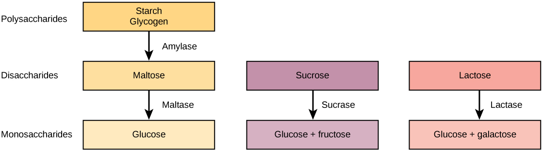
The enzyme amylase catalyzes the breakdown of food starches into maltose, a disaccharide. In many mammals, this enzymes is produced in saliva so carbohydrate breakdown starts in the mouth.
The disaccharides are then broken down into monosaccharides by enzymes called maltases, sucrases, and lactases. Maltase breaks down maltose into glucose. Other disaccharides, such as sucrose and lactose are broken down by sucrase and lactase, respectively. Sucrase breaks down sucrose (or “table sugar”) into glucose and fructose, and lactase breaks down lactose (or “milk sugar”) into glucose and galactose. The monosaccharides (e.g. glucose) thus produced are absorbed and then can be used in metabolic pathways to harness energy. The monosaccharides are transported across the gut epithelium so that all cells in the body can access them. In larger animals, the monosaccharides are moved into the bloodstream to be transported to the different cells in the body. The steps in carbohydrate digestion are summarized in Figure 5.1.1.
Practice Questions
Protein
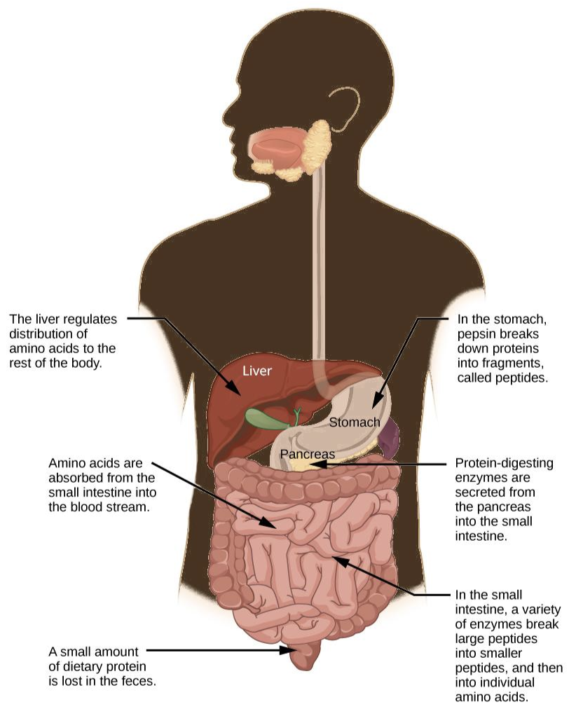
Proteases are enzymes that hydrolyze peptide bonds and break down proteins. In mammals, this begins in the stomach where the enzyme pepsin plays an important role in the digestion of proteins by breaking down the intact protein to peptides, which are short chains of four to nine amino acids. Further breakdown of peptides to single amino acids is aided by enzymes called peptidases (those that breakdown peptides). The amino acids are absorbed into the body through the epithelial lining of the gut. The steps in protein digestion in humans are summarized in Figure 5.1.2.
Practice Questions
Lipids
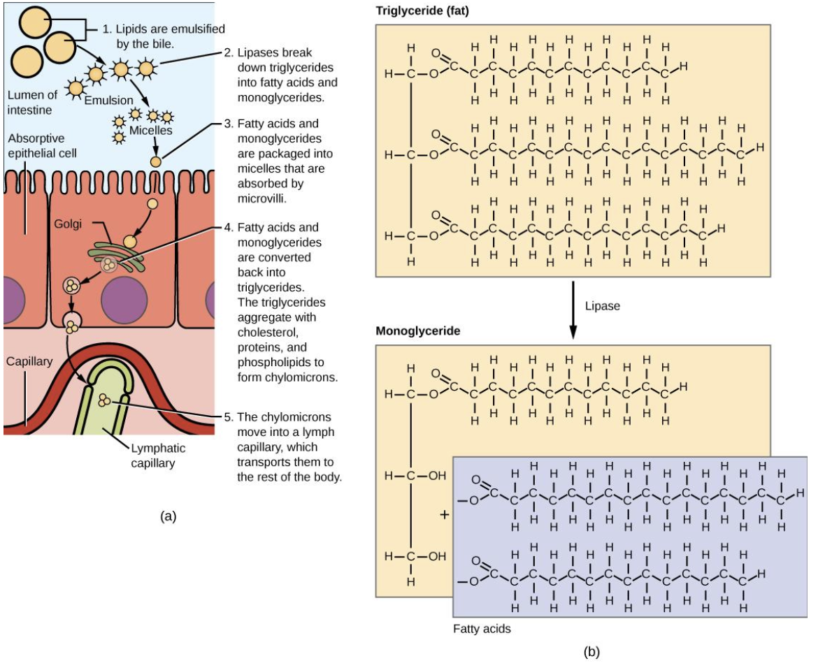
Lipases are enzymes that specialize in breaking down lipids. Lipids are hydrophobic substances: in the presence of water, they will aggregate to form globules to minimize exposure to water. This reduces the surface area of lipids that are available for the lipases to act on, leaving lipid digestion incomplete. Therefore, molecules that help in the process of emulsification are used to aid lipid digestion. Emulsification is a process in which large lipid globules are separated into several small lipid globules. By forming an emulsion, the available surface area of the lipids increases many fold. In mammals, this is accomplished by bile produced by the pancreas. Bile contains bile salts, which are amphipathic, meaning they contain hydrophobic and hydrophilic parts. Thus, the bile salts hydrophilic side can interface with water on one side and the hydrophobic side interfaces with lipids on the other. By doing so, bile salts emulsify large lipid globules into small lipid globules.
Lipases breakdown the lipids into fatty acids and glycerides (Figure 5.1.3). These molecules can pass through the plasma membrane of the cell and enter the epithelial cells of the intestinal lining. The bile salts surround long-chain fatty acids and monoglycerides forming tiny spheres called micelles. The micelles move into the brush border of the small intestine absorptive cells where the long-chain fatty acids and monoglycerides diffuse out of the micelles into the absorptive cells by simple diffusion leaving the micelles behind in the gut. The long-chain fatty acids and monoglycerides recombine in the absorptive cells to form triglycerides, which aggregate into globules and become coated with proteins. These large spheres are called chylomicrons. Chylomicrons contain triglycerides, cholesterol, and other lipids and have proteins on their surface. The surface is also composed of the hydrophilic phosphate “heads” of phospholipids. Together, they enable the chylomicron to move in an aqueous environment without exposing the lipids to water. Chylomicrons leave the absorptive cells via exocytosis. Chylomicrons ultimately enter the bloodstream where they can deliver triglycerides and cholesterol to the body’s tissues. They contribute to the production of high-density and low-density lipoproteins (HDL and LDL respectively) in the bloodstream.
Vitamins
Vitamins can be either water-soluble or lipid-soluble. Fat soluble vitamins are absorbed in the same manner as lipids. It is important to consume some amount of dietary lipid to aid the absorption of lipid-soluble vitamins. Water-soluble vitamins can be directly absorbed into the bloodstream from the gut.
Elimination
The final step in digestion is the elimination of undigested food content and waste products. Importantly, water is reabsorbed prior to the elimination.
Common Problems with Elimination
Diarrhea and constipation are some of the most common health concerns that affect digestion. Constipation is a condition where the feces are hardened because of excess water removal. In contrast, if enough water is not removed from the feces, it results in diarrhea. Many bacteria, including the ones that cause cholera, affect the proteins involved in water reabsorption in the colon and result in excessive diarrhea.
Emesis
Emesis, or vomiting, is elimination of food by forceful expulsion through the mouth. It is often in response to an irritant that affects the digestive tract, including but not limited to viruses, bacteria, emotions, sights, and food poisoning. This forceful expulsion of the food is due to the strong contractions produced by the stomach muscles and regulated by the medulla.
Practice Questions
Glossary
ingestion
the process of taking in food through the mouth
digestion
chemical breakdown of food into small organic fragments
absorption
the movement of monomers from the gut lumen into the bloodstream
elimination
excretion of undigested food and waste products from the body
Figure Descriptions
Figure 5.1.1. The figure is a three-path flowchart of carbohydrate digestion arranged from top to bottom by polymer size, with labels along the left margin for polysaccharides, disaccharides, and monosaccharides. In the first path, starch/glycogen is converted to maltose by amylase, and maltose is converted to glucose by maltase. In the second path, sucrose is split by sucrase into glucose and fructose. In the third path, lactose is split by lactase into glucose and galactose. Arrows point downward from substrate to enzyme to product, emphasizing that different enzymes target specific carbohydrates to yield monosaccharides. [Return to Figure 5.1.1]
Figure 5.1.2. A silhouette of a person shows the digestive organs with labels and arrows marking the steps of protein digestion. Food travels down the esophagus to the stomach, where pepsin begins digestion by cutting proteins into peptides. The pancreas releases protein-digesting enzymes into the small intestine, where multiple enzymes cleave large peptides into smaller peptides and finally into amino acids. These amino acids are absorbed from the small intestine into the bloodstream, and the liver regulates their distribution to the body. A note at the end of the tract indicates that a small amount of dietary protein is lost in the feces. Transcribed text: “In the stomach, pepsin breaks down proteins into fragments, called peptides.”; “Protein-digesting enzymes are secreted from the pancreas into the small intestine.”; “In the small intestine, a variety of enzymes break large peptides into smaller peptides, and then into amino acids.”; “Amino acids are absorbed from the small intestine into the blood stream.”; “The liver regulates distribution of amino acids to the rest of the body.”; “A small amount of dietary protein is lost in the feces.” [Return to Figure 5.1.2]
Figure 5.1.3. The image has two parts. On the left, there is a depiction of the steps of digestion through the small and large intestine. In Step 1, the image shows: in the intestinal lumen above the microvilli, large yellow fat droplets being broken into many small droplets by bile salts (emulsification). Transcribed text: “1. Lipids are emulsified by the bile.” In step 2, the image shows: purple lipase enzymes acting on the emulsified droplets, releasing small products. Transcribed text: “2. Lipases break down triglycerides into fatty acids and monoglycerides.” In step 3, the image shows: fatty acids and monoglycerides assembling into micelles near the brush border; arrows point to uptake at the microvilli of an absorptive epithelial cell. Transcribed text: “3. Fatty acids and monoglycerides are packaged into micelles and are absorbed by microvilli.” In step 4, the image shows: inside the epithelial cell, the absorbed products are re-esterified into triglycerides and packaged with cholesterol, proteins, and phospholipids into large lipoprotein spheres (chylomicrons) near the ER/Golgi. Transcribed text: “4. Fatty acids and monoglycerides are converted back into triglycerides. The triglycerides aggregate with cholesterol, proteins, and phospholipids to form chylomicrons.” In step 5, the image shows: chylomicrons exiting the cell’s basal side and entering a green lymphatic capillary (lacteal) next to a blood capillary. Transcribed text: “5. The chylomicrons move into a lymph capillary, which transports them to the rest of the body.” On the right, there is an image of a chemical close-up: The image shows a beige box labeled Triglyceride (fat) split by an arrow labeled Lipase into a beige Monoglyceride and a blue panel of Fatty acids. Transcribed labels: “Triglyceride (fat),” “Lipase,” “Monoglyceride,” “Fatty acids.” [Return to Figure 5.1.3]
Licenses and Attributions
“5.1 Digestive Processes in Animals” is adapted from “34.3 Digestive System Processes” by Mary Ann Clark, Matthew Douglas, and Jung Choi for OpenStax Biology 2e under CC-BY 4.0. “5.1 Digestive Processes in Animals” is licensed under CC-BY-NC 4.0.
Media Attributions
- 1A.B.digestion of carbs
- 1A.B.digestion of proteins
- 1A.B.lipid digestion and absorption
definition
the process of taking in food through the mouth
chemical breakdown of food into small organic fragments
the movement of monomers from the gut lumen into the bloodstream
excretion of undigested food and waste products from the body
