17.2 Organization of DNA Inside a Cell
Elizabeth Dahlhoff
Learning Objectives
By the end of this section, you will be able to do the following:
- Describe the organization of DNA in prokaryotic cells, including the structure and location of the genome and plasmids.
- Compare and contrast the structure and organization of prokaryotic and eukaryotic genomes.
- Differentiate between haploid and diploid cells.
- Describe the role of histone proteins and nucleosomes in eukaryotic DNA compaction.
- Explain how genetic variation arises from different combinations of alleles inherited from each parent.
DNA Organization in Prokaryotes
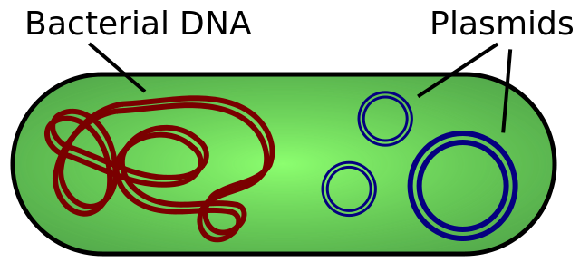
All of a cell’s DNA, packaged as a double-stranded DNA molecule, is called its genome. In prokaryotes, the genome is composed of a single, double-stranded DNA molecule in the form of a loop or circle (Figure 17.2.1). The region in the cell containing this genetic material is called a nucleoid (remember that prokaryotes do not have a separate membrane-bound nucleus). Some prokaryotes also have smaller loops of DNA called plasmids that are not essential for normal growth. Bacteria can exchange these plasmids with other bacteria, sometimes receiving beneficial new genes that the recipient can add to their chromosomal DNA. Antibiotic resistance is one trait that often spreads through a bacterial colony through plasmid exchange.
The size of the genome in one of the most well-studied prokaryotes, E.coli, is 4.6 million base pairs (which would be approximately 1.1 mm in length, if stretched out end to end). So how does this fit inside a small bacterial cell? The DNA is twisted by what is known as supercoiling. Supercoiled DNA is coiled more tightly than would be typically be found in a cell (more than 10 nucleotides per twist of the helix). If you visualize twisting a rope until it twists back on itself, you have a pretty good visual of supercoiled DNA. This process allows the DNA to be compacted into the small space inside a bacteria.
DNA Organization in Eukaryotes
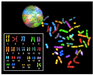
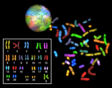
Eukaryotes have much more DNA than prokaryotes. For example, an E. coli bacteria contains roughly 3 million base pairs of DNA, while a human contains roughly 3 billion. In eukaryotes such as humans and other animals, the genome consists of several double-stranded linear DNA molecules (Figure 17.2.2), which are located inside a membrane-bound nucleus. Each species of eukaryotes has a characteristic number of chromosomes in the nuclei (plural of nucleus) of its cells. A normal human gamete (sperm or egg) contains 23 chromosomes. A normal human body cell, or somatic cell, contains 46 chromosomes (one set of 23 from the egg and one set of 23 from the sperm; Figure 17.2.2). The letter n is used to represent a single set of chromosomes; therefore, a gamete (sperm or egg) is designated 1n, and is called a haploid cell. Somatic cells (body cells) are designated 2n and are called diploid cells.
Matched pairs of chromosomes in a diploid organism are called homologous (“same knowledge”) chromosomes. Of a pair of homologous chromosomes, one came from the egg and the second came from the sperm. Homologous chromosomes are the same length and have specific nucleotide segments called genes in exactly the same location, or locus (plural = loci). Genes, the functional units of chromosomes, determine specific characteristics by coding for specific proteins. Traits are the variations of those characteristics. For example, we remember from our reading about Mendel that pea flower color is a characteristic, with traits that are violet or white.
Each copy of a homologous pair of chromosomes originates from a different parent; therefore, the sequence of DNA present in the two genes on a pair of homologous chromosomes is not necessarily identical, despite the same genes being present in the same locations on the chromosome. These different versions of a gene that contain different sequences of DNA are called alleles.
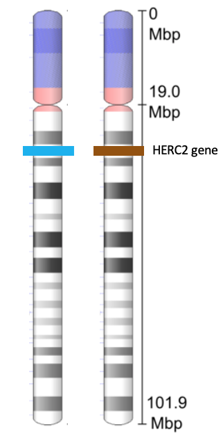
If you look at Figure 17.2.3, you can see a pair of homologous chromosomes. The chromosome shown in the figure is human chromosome 15. The HERC2 gene is located on this chromosome. The HERC2 gene is one of at least three genes involved in determining human eye color. The HERC2 gene is also involved in DNA repair, centrosome assembly, iron metabolism, and is associated with some genetic diseases (Sánchez-Tena et al., 2016). Each person inherits two copies of the HERC2 gene: one from the egg and one from the sperm. However, the alleles of the HERC2 gene that they inherit can be different. In the figure, the cell containing this homologous pair of chromosomes contains one blue allele and one brown allele.
The variation of individuals within a species is due to the specific combination of the genes inherited from both parents. Even a slightly altered sequence of nucleotides within a gene can result in an alternative trait. For example, we have learned (Chapter 15) that there are three possible gene sequences (alleles) on the human chromosome that code for blood type: sequence A, sequence B, and sequence O. Because all diploid human cells have two copies of the chromosome that determines blood type, the blood type (the trait) is determined by which two versions of the marker gene are inherited. It is possible to have two copies of the same allele on both homologous chromosomes, with one on each (for example, AA, BB, or OO; homozygous), or two different alleles, such as AB (heterozygous).
Minor variations of traits, such as flower color, blood type, eye color, and handedness, contribute to the natural variation found within a species. However, if the entire DNA sequence from any pair of homologous chromosomes is compared, the difference is less than 1%. The sex chromosomes, (X and Y in humans), are the single exception to the rule of homologous chromosome uniformity: Other than a small amount of homology that is necessary to accurately produce gametes, the genes found on the X and Y chromosomes are different.
Eukaryotic Chromosomal Structure and Compaction
Continuing with humans as our example, if the DNA from all 46 chromosomes in a human cell nucleus was laid out end to end, it would measure approximately two meters; however, its diameter would be only 2 nm. Considering that the size of a typical human cell is about 10 µm (100,000 cells lined up to equal one meter), DNA must be tightly packaged to fit in the cell’s nucleus. At the same time, it must also be readily accessible for genes to be expressed. During some stages of the cell cycle, the long strands of DNA are condensed into compact chromosomes.
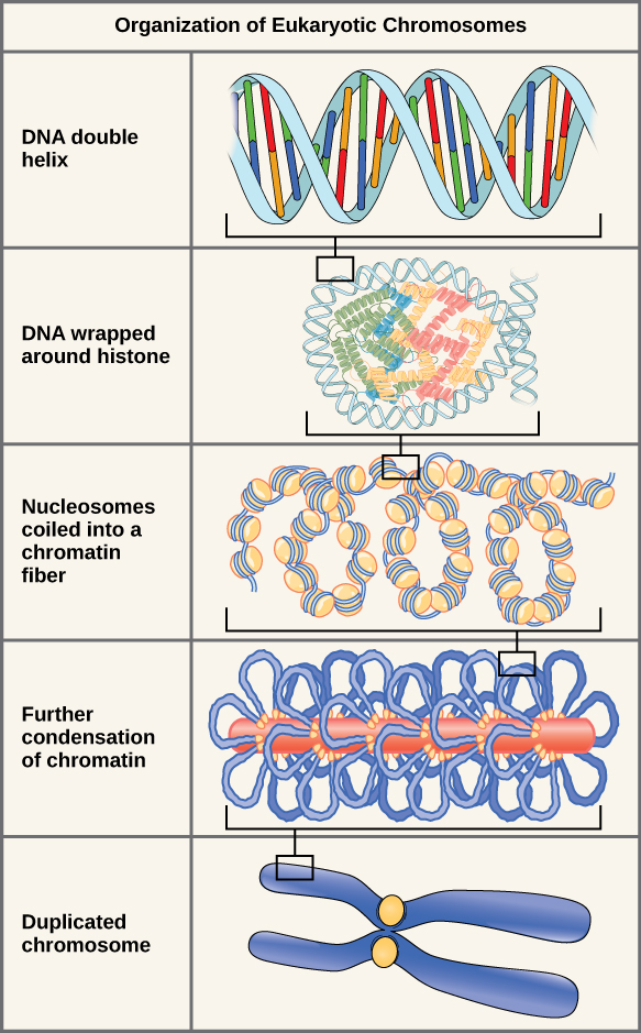
Eukaryotes, whose chromosomes each consist of a linear DNA molecule, employ a complex type of packing strategy to fit their DNA inside the nucleus (Figure 17.2.4). At the most basic level, DNA is wrapped around proteins known as histones to form structures called nucleosomes. The histones are evolutionarily conserved proteins that form an octamer of eight histone proteins attached together. DNA, which is negatively charged because of the phosphate groups, is wrapped tightly around the histone core, which has an overall positive charge. This nucleosome is linked to the next one with the help of a linker DNA. This is also known as the “beads on a string” structure. This is further compacted into a 30 nm fiber, which is the diameter of the structure. At the metaphase stage, the chromosomes are at their most compact, are approximately 700 nm in width, and are found in association with scaffold proteins.
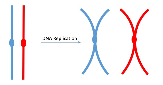
DNA replicates in the S phase of interphase. After replication, the chromosomes are composed of two linked sister chromatids (Figure 17.2.5). This means that the only time chromosomes look like an “X” is after DNA replication has taken place and the chromosomes have condensed. During the majority of the cell’s life, chromosomes are composed of only one copy and they are not tightly compacted into chromosomes. When fully compact, the pairs of identically packed chromosomes are bound to each other by cohesin proteins. The connection between the sister chromatids is closest in a region called the centromere. The conjoined sister chromatids, with a diameter of about 1 µm, are visible under a light microscope. The centromeric region is highly condensed and thus will appear as a constricted area. In Figure 17.2.4, it is shown as an oval because it is easier to draw that way.
Section Summary
In all cells, DNA is organized to fit within the limited space of the cell. In prokaryotes, the genome is a single circular DNA molecule located in the nucleoid region. These cells may also contain plasmids, which are small, circular DNA molecules that can carry beneficial genes such as those for antibiotic resistance. To fit inside the small cell, prokaryotic DNA is tightly packed through supercoiling. Eukaryotes have larger, linear genomes housed within a membrane-bound nucleus. Somatic cells have diploid or 2n chromosomes, while gametes (sperm and egg) have haploid or 1n chromosomes. Chromosomes come in homologous pairs, one from each parent, and carry genes at the same loci. Different versions of a gene, called alleles, can produce different traits. To compact their large genomes, eukaryotes wrap DNA around histone proteins to form nucleosomes, creating a “beads on a string” structure. These nucleosomes are further coiled into thicker fibers and compacted even more during cell division. After DNA replication, each chromosome consists of two identical sister chromatids joined at a centromere. This compact structure ensures DNA fits in the nucleus while remaining accessible for gene expression and replication.
Practice Questions
Glossary
- allele
one of multiple possible versions of a gene found at the same location (locus) on homologous chromosomes; alleles may differ slightly in their DNA sequence and result in different traits - chromatin
complex of DNA and protein (primarily histones) found in eukaryotic nuclei; chromatin condenses to form visible chromosomes during cell division - chromosome
single, continuous DNA molecule with associated proteins, carrying genetic information; eukaryotes have linear chromosomes, while prokaryotes have a circular chromosome - diploid (2n)
a cell that contains two complete sets of chromosomes, one from each parent; most human somatic cells are diploid, with 46 chromosomes total - gamete
haploid reproductive cell (sperm or egg) that carries one set of chromosomes (n); fusion of two gametes forms a diploid zygote - gene
segment of DNA located on a chromosome that codes for a specific protein or RNA product, contributing to the inheritance of a particular trait
haploid (n) - cell that contains one set of chromosomes; human gametes (sperm and eggs) are haploid
- histone
positively charged protein that helps package and order DNA into structural units called nucleosomes in eukaryotic cells - homologous chromosomes
matched pair of chromosomes in a diploid organism, one inherited from each parent, with same genes in the same order but possibly different alleles - locus
specific physical location of a gene on a chromosome (plural = loci) - sister chromatids
two identical copies of a chromosome that are connected at the centromere following DNA replication - trait
specific variation of a characteristic (such as blue versus brown eyes) determined by different alleles of a gene
Figure Descriptions
Figure 17.2.1. The image is a simplified diagram of a prokaryotic cell. It has an oval shape with a light green background representing the cytoplasm. Inside, there are several key components labeled with black text. To the left, there is a red circular and looped structure representing the genomic DNA. On the right side, there are three concentric circles in blue indicating plasmids. The cell is outlined with a black border, and external features like the cell wall and plasmids are labeled with black lines pointing from the text to the components. [Return to Figure 17.2.1]
Figure 17.2.2. The image shows three views of chromosomes from a female human somatic cell. At the top, condensed chromosomes are visible inside a colorful, round nucleus. On the right, the same chromosomes, removed from a cell in mitosis, are spread out and stained with fluorescent dyes, each chromosome displaying a different color. On the left, the chromosomes are artificially arranged in numbered pairs from 1 to 22 plus the X chromosome, creating a karyotype. This chromosome painting technique uses fluorescent stains to differentiate chromosomes by color. [Return to Figure 17.2.2]
Figure 17.2.3. The image shows two vertically oriented chromosome 15s side by side, with banding patterns in shades of gray. A vertical scale marks positions from 0 to 101.9 megabase pairs (Mbp), with 19.0 Mbp labeled partway down. On the left chromosome, a horizontal blue bar marks one allele of the HERC2 gene, while on the right chromosome, a horizontal brown bar marks the other allele. The blue and brown coloring represents two different alleles of the HERC2 gene. [Return to Figure 17.2.3]
Figure 17.2.4. The diagram, titled Organization of Eukaryotic Chromosomes, illustrates the progressive packaging of DNA into a duplicated chromosome in five stages. At the top, the “DNA double helix” is shown as a blue twisted ladder with multicolored base pairs. Below, “DNA wrapped around histone” depicts the helix coiled around histone proteins, forming bead-like nucleosomes. The next stage, “Nucleosomes coiled into a chromatin fiber,” shows these beads twisted into a thicker strand. In “Further condensation of chromatin,” the fiber is looped and compacted into an even denser arrangement. Finally, “Duplicated chromosome” shows two identical blue sister chromatids connected at a yellow centromere. Black brackets and magnification indicators connect the stages, demonstrating the increasing levels of DNA condensation. [Return to Figure 17.2.4]
Licenses and Attributions
This chapter, “Organization of DNA Inside a Cell,” by Elizabeth Dahlhoff, is adapted from “DNA Organization inside a cell”from in Principles of Biology by Lisa Bartree, Walter Shriner and Catherine Creech (Mt. Hood Community College) under a CC BY-NC 4.0 license. This work is licensed under a CC BY-NC 4.0 license.
Media Attributions
- 1B.17.2.ProkaryoticDNA © Spaully is licensed under a CC BY-SA (Attribution ShareAlike) license
- euk DNA org

.svg)