17.4 DNA Repair
Elizabeth Dahlhoff
Learning Objectives
By the end of this section, you will be able to do the following:
- Describe the different types of errors that can occur during DNA replication.
- Explain DNA repair mechanisms.
DNA Repair
DNA replication is a highly accurate process, but mistakes can occasionally occur, such as a DNA polymerase inserting a wrong base. Uncorrected mistakes may sometimes lead to serious consequences, such as cancer. Repair mechanisms correct the mistakes. In rare cases, mistakes are not corrected, leading to mutations; in other cases, repair enzymes are themselves mutated or defective.
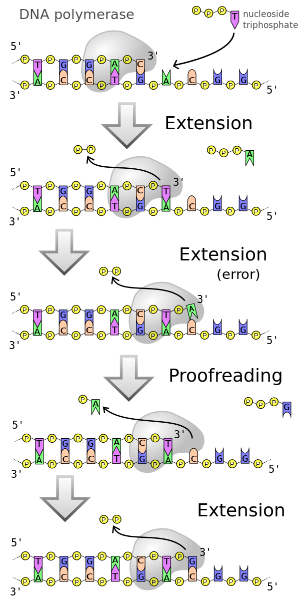
Most of the mistakes during DNA replication are promptly corrected by DNA polymerase by proofreading the base that has been just added (Figure 17.4.1). In proofreading, the DNA pol reads the newly added base before adding the next one, so a correction can be made. The polymerase checks whether the newly added base has paired correctly with the base in the template strand. If it is the right base, the next nucleotide is added. If an incorrect base has been added, the enzyme makes a cut at the phosphodiester bond and releases the wrong nucleotide. This is performed by the exonuclease action of DNA pol III. Once the incorrect nucleotide has been removed, a new one will be added again (Figure 17.4.1).
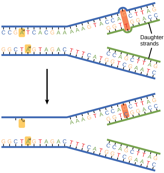
Some errors are not corrected during replication but are instead corrected after replication is completed; this type of repair is known as mismatch repair (Figure 17.4.2). The enzymes recognize the incorrectly added nucleotide and excise it; this is then replaced by the correct base. If this remains uncorrected, it may lead to more permanent damage. How do mismatch repair enzymes recognize which of the two bases is the incorrect one? In E. coli, after replication, the nitrogenous base adenine acquires a methyl group (CH3); the parental DNA strand will have methyl groups, whereas the newly synthesized strand lacks them. Thus, DNA polymerase is able to remove the wrongly incorporated bases from the newly synthesized, non-methylated strand. In eukaryotes, the mechanism is not very well understood, but it is believed to involve recognition of unsealed nicks in the new strand, as well as a short-term continuing association of some of the replication proteins with the new daughter strand after replication has completed.
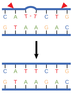
In another type of repair mechanism, nucleotide excision repair, enzymes replace incorrect bases by making a cut on both the 3′ and 5′ ends of the incorrect base (Figure 17.4.3). The segment of DNA is removed and replaced with the correctly paired nucleotides by the action of DNA pol. Once the bases are filled in, the remaining gap is sealed with a phosphodiester linkage catalyzed by DNA ligase. This repair mechanism is often employed when UV exposure causes the formation of thymine-thymine dimers (the small – connecting the two Ts in Figure 17.4.3).
A well-studied example of mistakes not being corrected is seen in people suffering from xeroderma pigmentosa. Affected individuals have skin that is highly sensitive to UV rays from the sun. When individuals are exposed to UV, pyrimidine dimers, especially those of thymine, are formed; people with xeroderma pigmentosa are not able to repair the damage. These are not repaired because of a defect in the nucleotide excision repair enzymes, whereas in normal individuals, the thymine dimers are excised and the defect is corrected. The thymine dimers distort the structure of the DNA double helix, and this may cause problems during DNA replication. People with xeroderma pigmentosa have a higher risk of contracting skin cancer than those who don’t have the condition.
Errors during DNA replication are not the only reason why mutations arise in DNA. Mutations, variations in the nucleotide sequence of a genome, can also occur because of damage to DNA. Such mutations may be of two types: induced or spontaneous. Induced mutations are those that result from an exposure to a mutagen: chemicals, UV rays, X-rays, or some other environmental agent. Spontaneous mutations occur without any exposure to any environmental agent; they are a result of natural reactions taking place within the body. Mutations may have a wide range of effects. Some mutations are not expressed; these are known as silent mutations. Other mutations can have serious effects on the organism (such as the mutation that causes xeroderma pigmentosa.
Mutations in repair genes have been known to cause cancer. Many mutated repair genes have been implicated in certain forms of pancreatic cancer, colon cancer, and colorectal cancer. Mutations can affect either somatic cells or gametes. If many mutations accumulate in a somatic cell, they may lead to problems such as the uncontrolled cell division observed in cancer. If a mutation takes place in a gamete, the mutation can be passed on to the next generation.
Practice Questions
Section Summary
DNA polymerase can make mistakes while adding nucleotides. It edits the DNA by proofreading every newly added base. Incorrect bases are removed and replaced by the correct base, and then a new base is added. Most mistakes are corrected during replication, although when this does not happen, the mismatch repair mechanism is employed. Mismatch repair enzymes recognize the wrongly incorporated base and excise it from the DNA, replacing it with the correct base. In yet another type of repair, nucleotide excision repair, the incorrect base is removed along with a few bases on the 5′ and 3′ end, and these are replaced by copying the template with the help of DNA polymerase. The ends of the newly synthesized fragment are attached to the rest of the DNA using DNA ligase, which creates a phosphodiester bond. Most mistakes are corrected, and if they are not, they may result in a mutation defined as a permanent change in the DNA sequence. Mutations can be of many types, such as substitution, deletion, insertion, and translocation. Mutations in repair genes may lead to serious consequences such as cancer. Mutations can be induced or may occur spontaneously.
Glossary
- induced mutation
- mutation that results from exposure to chemicals or environmental agents
- mutation
- variation in the nucleotide sequence of a genome
- mismatch repair
- type of repair mechanism in which mismatched bases are removed after replication
- nucleotide excision repair
- type of DNA repair mechanism in which the wrong base, along with a few nucleotides upstream or downstream, are removed
- proofreading
- function of DNA pol in which it reads the newly added base before adding the next one
- point mutation
- mutation that affects a single base
- silent mutation
- mutation that is not expressed
- spontaneous mutation
- mutation that takes place in the cells as a result of chemical reactions taking place naturally without exposure to any external agent
- transition substitution
- when a purine is replaced with a purine or a pyrimidine is replaced with another pyrimidine
- transversion substitution
- when a purine is replaced by a pyrimidine or a pyrimidine is replaced by a purine
Figure Descriptions
Figure 17.4.1. The figure, titled “DNA polymerase,” shows a five-step vertical schematic illustrating proofreading during DNA replication. At the top right, a key identifies “nucleoside triphosphate” with a purple T-shaped icon, and numerous yellow circles marked P represent phosphates. In each panel, a gray enzyme labeled “3′” is positioned on a DNA duplex with bases labeled “A,” “T,” “G,” and “C,” while large white arrows point downward between panels to show progression. The first panel, labeled “Extension,” shows DNA polymerase adding nucleotides to a growing strand complementary to the template. The second panel, labeled “Extension (error),” depicts the addition of an incorrect base, creating a mismatch. The third panel, labeled “Proofreading,” shows the polymerase pausing to remove the incorrect nucleotide. The fourth panel, labeled “Extension,” depicts the correct nucleoside triphosphate being added. The fifth panel shows the completed DNA duplex with all base pairs correctly matched. [Return to Figure 17.4.1]
Figure 17.4.2. The diagram illustrates mismatch repair in three stages. At the top, two daughter DNA strands are shown after replication, with one containing an incorrect base pair—an adenine (A) mismatched with cytosine (C). A section of one strand is labeled “Daughter strands.” In the middle panel, an arrow points downward to a DNA segment where mismatch repair proteins have detected the error; the mismatched base is highlighted, and an enzyme is shown removing it from the newly synthesized strand. In the bottom panel, the gap has been filled with the correct base to restore proper base pairing, completing the repair process. [Return to Figure 17.4.2]
Figure 17.4.3. The image shows two steps of nucleotide excision repair. In the first panel, a DNA double strand contains two adjacent thymine bases on the top strand bonded together, forming a thymine dimer, represented by a bulge between them. Red arrowheads mark the start and end points where the damaged section will be cut out. In the second panel, the thymine dimer is removed, and the DNA sequence is restored to its correct form, with complementary base pairing between the top and bottom strands. [Return to Figure 17.4.3]
Licenses and Attributions
This chapter, “DNA Repair,” by Elizabeth Dahlhoff, is adapted from chapters in the “DNA Structure and Function” section in Biology by Open Stax College (University of Hawaii) under a CC BY-NC 4.0 license. This work is licensed under a CC BY-NC 4.0 license.
Media Attributions
- 600px-DNA_polymerase
- Figure_14_06_02
- Figure_14_06_03
