17.3 DNA Replication
Elizabeth Dahlhoff
Learning Objectives
By the end of this section, you will be able to do the following:
- Explain the process of DNA replication in prokaryotes.
- Describe the role of different enzymes and proteins in supporting this process.
- Compare and contrast DNA replication in eukaryotes and prokaryotes.
- Explain the role of telomerase in DNA replication.
DNA Replication in Prokaryotes
DNA replication has been extremely well studied in prokaryotes primarily because of the small size of the genome and the mutants that are available. E. coli has 4.6 million base pairs in a single circular chromosome and all of it gets replicated in approximately 42 minutes, starting from a single origin of replication and proceeding around the circle in both directions. This means that approximately 1,000 nucleotides are added per second. The process is quite rapid and occurs without many mistakes.
DNA replication employs a large number of proteins and enzymes, each of which plays a critical role during the process. One of the key players is the enzyme DNA polymerase, also known as DNA pol, which adds nucleotides one by one to the growing DNA chain that are complementary to the template strand. The addition of nucleotides requires energy; this energy is obtained from the nucleotides that have three phosphates attached to them, similar to ATP which has three phosphate groups attached. When the bond between the phosphates is broken, the energy released is used to form the phosphodiester bond between the incoming nucleotide and the growing chain. In prokaryotes, three main types of polymerases are known: DNA pol I, DNA pol II, and DNA pol III. It is now known that DNA pol III is the enzyme required for DNA synthesis; DNA pol I and DNA pol II are primarily required for repair.
How does the replication machinery know where to begin? It turns out that there are specific nucleotide sequences called origins of replication where replication begins. In E. coli, which has a single origin of replication on its one chromosome (as do most prokaryotes), it is approximately 245 base pairs long and is rich in AT sequences. The origin of replication is recognized by certain proteins that bind to this site.
An enzyme called helicase unwinds the DNA by breaking the hydrogen bonds between the nitrogenous base pairs. ATP hydrolysis is required for this process. As the DNA opens up, Y-shaped structures called replication forks are formed. Two replication forks (a replication bubble) are formed at the origin of replication and these get extended bi- directionally as replication proceeds. Single-strand binding proteins coat the single strands of DNA near the replication fork to prevent the single-stranded DNA from winding back into a double helix. DNA polymerase is able to add nucleotides only in the 5′ to 3′ direction (a new DNA strand can be only extended in this direction). It also requires a free 3′-OH group to which it can add nucleotides by forming a phosphodiester bond between the 3′-OH end and the 5′ phosphate of the next nucleotide. This essentially means that it cannot add nucleotides if a free 3′-OH group is not available. Then how does it add the first nucleotide? The problem is solved with the help of a primer that provides the free 3′-OH end.
Another enzyme, RNA primase, synthesizes an RNA primer that is about five to ten nucleotides long and complementary to the DNA. Because this sequence primes the DNA synthesis, it is appropriately called the primer. DNA polymerase can now extend this RNA primer, adding nucleotides one by one that are complementary to the template strand.
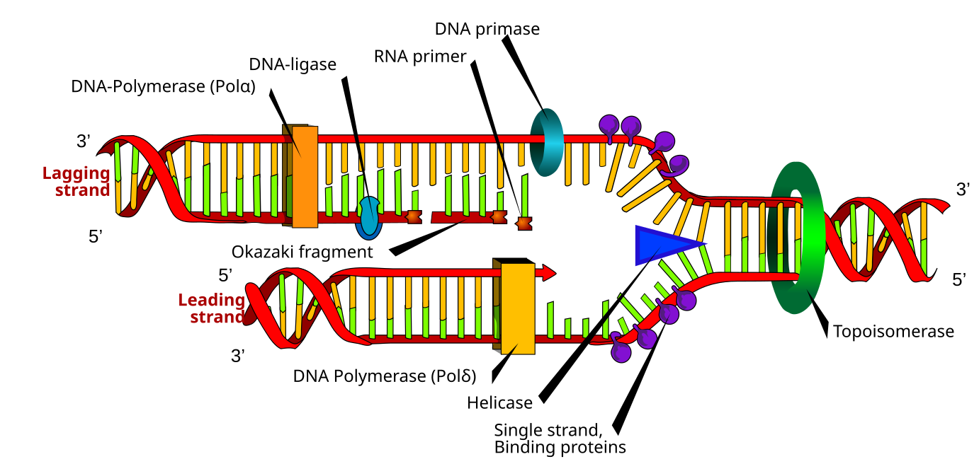
Because DNA polymerase can only extend in the 5′ to 3′ direction, and because the DNA double helix is antiparallel, there is a challenge or obstacle at the replication fork. Since the two template DNA strands have opposing orientations: only one new DNA strand can be synthesized continuously towards the replication fork. This continuously synthesized strand is known as the leading strand. The other strand is extended away from the replication fork, and is synthesized in numerous small fragments known as Okazaki fragments, each requiring a primer to start the synthesis (Figure 17.3.1). New primer segments are laid down in the direction of the replication fork, but each pointing away from it. Okazaki fragments are named after the Japanese scientist who first discovered them. The strand with the Okazaki fragments is known as the lagging strand. The leading strand can be extended from a single primer, whereas the lagging strand needs a new primer for each of the short Okazaki fragments. The overall direction of the lagging strand will be 3′ to 5′, and that of the leading strand 5′ to 3′.
As synthesis proceeds, the RNA primers are replaced by DNA. In E. coli, the removal of RNA nucleotides and their replacement with DNA nucleotides are both performed by the enzyme DNA polymerase I. The nick that remains between the newly synthesized DNA (that replaced the RNA primer) and the previously synthesized DNA are sealed by the enzyme DNA ligase, which catalyzes the formation of the phosphodiester linkage between the 3′-OH end of one nucleotide and the 5′ phosphate end of the other fragment.
After the chromosome has been completely replicated, the two DNA copies will be moved into two different cells during cell division.
DNA Replication is Semiconservative
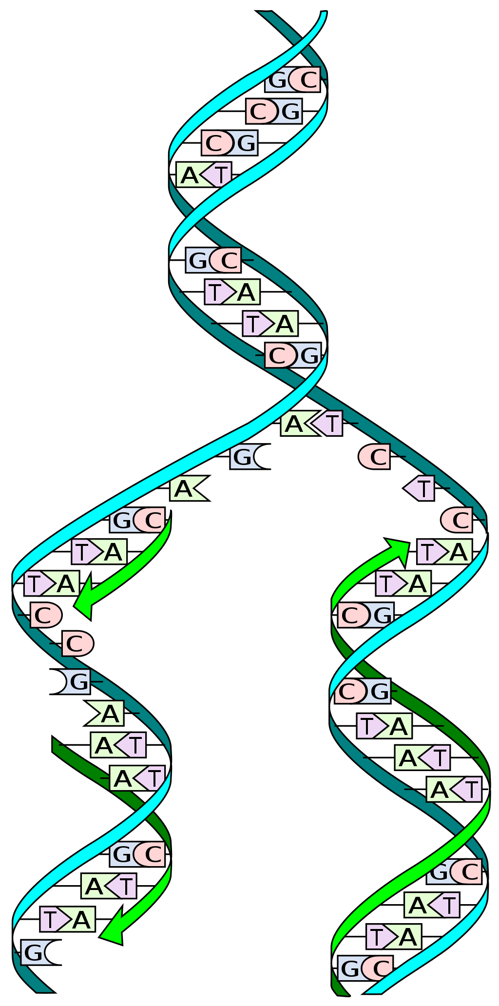
During DNA replication, each of the two strands that make up the double helix serves as a template from which a new strand is copied. The new strands will be complementary to the parental or “old” strands. When two daughter DNA copies are formed, they have the same sequence and are divided equally into the two daughter cells.
Eukaryotic DNA Replication
Eukaryotic genomes are large and complex, and replication of them required multiple origins of replication. In addition, DNA replication is much slower than in prokaryotes and involves several specialized DNA polymerases to complete the process. Because of how eukaryotic chromosomes are packaged inside the nucleus as chromatin, this structure must be loosened to allow access. However, DNA replication follows the same basic steps in eukaryotes as it does in prokaryotes: unwinding DNA, primer synthesis, strand elongation, and ligation.
One major difference between prokaryotes and eukaryotes is that eukaryotic chromosomes are linear. This means that their ends, which are called telomeres, cannot be fully replicated on the lagging strand. Telomeres are made of repeating sequences that protect genes closest to the end of the chromosome from being lost or damaged. The enzyme telomerase, active in germ and stem cells, extends these ends using an RNA template. Interestingly, telomere shortening is linked to aging; telomerase reactivation in mice has shown potential to reverse age-related decline. In contrast, telomerase is much more active in cancer cells than normal cells. Cancer is characterized by uncontrolled cell division of abnormal cells. The cells accumulate mutations, proliferate uncontrollably, and can migrate to different parts of the body through a process called metastasis. Scientists have observed that cancerous cells have considerably shortened telomeres. Only after the telomeres were shortened in the cancer cells does telomerase become active. If the action of telomerase in these cells can be inhibited by drugs during cancer therapy, then the cancerous cells could potentially be stopped from further division.
| Property | Prokaryotes | Eukaryotes |
|---|---|---|
| Origin of replication | Single | Multiple |
| Rate of replication | 1,000 nucleotides | 50 to 100 nucleotides |
| DNA polymerase types | 5 | 14 |
| Telomerase | Not present | Present |
| RNA primer removal | DNA pol I | RNase H |
| Strand elongation | DNA pol III | Pol δ, pol ε |
| Sliding clamp | Sliding clamp | PCNA |
Here is an animated explanation of DNA replication.
Video 17.3.1. DNA replication – 3D by yourgenome
Practice Questions
Section Summary
Replication in prokaryotes starts from a sequence found on the chromosome called the origin of replication—the point at which the DNA opens up. Helicase opens up the DNA double helix, resulting in the formation of the replication fork. Single-strand binding proteins bind to the single-stranded DNA near the replication fork to keep the fork open. Primase synthesizes an RNA primer to initiate synthesis by DNA polymerase, which can add nucleotides only in the 5′ to 3′ direction. One strand is synthesized continuously in the direction of the replication fork; this is called the leading strand. The other strand is synthesized in a direction away from the replication fork, in short stretches of DNA known as Okazaki fragments. This strand is known as the lagging strand. Once replication is completed, the RNA primers are replaced by DNA nucleotides and the DNA is sealed with DNA ligase, which creates phosphodiester bonds between the 3′-OH of one end and the 5′ phosphate of the other strand. Replication in eukaryotes starts at multiple origins of replication, but otherwise the mechanism is quite similar to prokaryotes. A primer is required to initiate synthesis, which is then extended by DNA polymerase as it adds nucleotides one by one to the growing chain. The leading strand is synthesized continuously, whereas the lagging strand is synthesized in Okazaki fragments. The RNA primers are replaced with DNA nucleotides; the DNA remains one continuous strand by linking the DNA fragments with DNA ligase. The ends of the eukaryotic chromosomes pose a problem as polymerase is unable to extend them without a primer. Telomerase, an enzyme with an inbuilt RNA template, extends the ends by copying the RNA template and extending one end of the chromosome. DNA polymerase can then extend the DNA using the primer. In this way, the ends of the chromosomes are protected.
Glossary
- helicase
- during replication, this enzyme helps to open up the DNA helix by breaking the hydrogen bonds
- lagging strand
- during replication, the strand that is replicated in short fragments and away from the replication fork
- leading strand
- strand that is synthesized continuously in the 5′ to 3′ direction which is synthesized in the direction of the replication fork
- ligase
- enzyme that catalyzes the formation of a phosphodiester linkage between the 3′ OH and 5′ phosphate ends of the DNA
- Okazaki fragment
- DNA fragment that is synthesized in short stretches on the lagging strand
- primase
- enzyme that synthesizes the RNA primer; the primer is needed for DNA pol to start synthesis of a new DNA strand
- primer
- short stretch of nucleotides that is required to initiate replication; in the case of replication, the primer has RNA nucleotides
- replication bubble
- unwound and open region of DNA where DNA replication occurs. It’s formed when the enzyme helicase separates the two strands of DNA, creating a “bubble” shape where replication can proceed. Within the bubble, two replication forks are formed.
- replication fork
- Y-shaped structure formed during initiation of replication
- single-strand binding protein
- during replication, protein that binds to the single-stranded DNA; this helps in keeping the two strands of DNA apart so that they may serve as templates
- sliding clamp
- ring-shaped protein that holds the DNA pol on the DNA strand
- telomerase
- enzyme that contains a catalytic part and an inbuilt RNA template; it functions to maintain telomeres at chromosome ends
- telomere
- DNA at the end of linear chromosomes
topoisomerase
enzyme that causes under-winding or overwinding of DNA when DNA replication is taking place
Figure Descriptions
Figure 17.3.1. The image illustrates the process of DNA replication, focusing on the formation of new DNA strands. The diagram shows two strands: the leading strand and the lagging strand. The leading strand is synthesized continuously, depicted with a red and green color coil. The lagging strand, shown above the leading strand, is synthesized in short segments called Okazaki fragments, and is indicated by similar colors with a depiction of segments. Various enzymes and proteins are represented, such as helicase, polymerase, and primase, shown as different colored shapes interacting with the DNA strands. [Return to Figure 17.3.1]
Figure 17.3.2. The diagram shows a DNA double helix with one light blue strand and one dark blue strand. The helix is unwinding, and each old strand is used as a template to build a new strand, indicated by green arrows. Base pairs are labeled with the letters A, T, G, and C to show complementary pairing (A with T, G with C). After replication, two double helices form, each containing one old strand and one new strand. [Return to Figure 17.3.2]
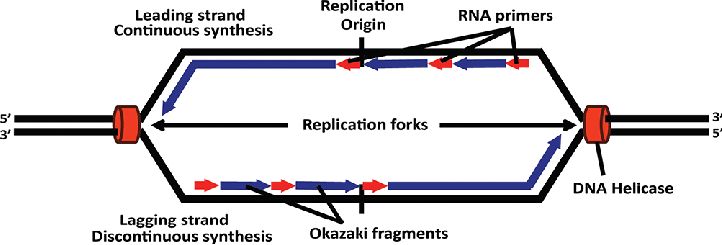
Figure 17.3.3. The diagram shows a “replication bubble” where DNA is being copied starting at the Replication Origin. Two Replication forks move in opposite directions. On each fork, the Leading strand is made in one smooth piece (Continuous synthesis) toward the fork, while the Lagging strand is made in short pieces (Discontinuous synthesis) called Okazaki fragments. These short pieces start with RNA primers (shown as red arrows). DNA Helicase (red ovals) unwinds the DNA strands at each fork. The DNA strands are marked with 5′ and 3′ labels to show direction. [Return to Figure 17.3.3]
Licenses and Attributions
Media Attributions
- 1B.17.3ReplicationFork © Mariana Ruiz Villareal is licensed under a Public Domain license
- DNA replication © Madeleine Price Ball is licensed under a CC0 (Creative Commons Zero) license
- Origin-of-bidirectional-replication
