14.6 The Eukaryotic Cell Cycle
Elizabeth Dahlhoff
Learning Objectives
By the end of this section, you will be able to do the following:
- Describe the three stages of interphase.
- Review the basic features of chromosomal division and cytokinesis during mitosis.
- Define the quiescent G0 phase.
Introduction
Eukaryotes have two major types of cell division: mitosis and meiosis. Mitosis is used to produce new body cells for growth and healing, while meiosis is used to produce sex cells (eggs and sperm). Meiosis will be discussed in a later chapter.
The cell cycle is an ordered series of events involving cell growth and cell division that produces two new daughter cells via mitosis. The length of the cell cycle is highly variable even within the cells of an individual organism. In humans, the frequency of cell turnover ranges from a few hours in early embryonic development to an average of two to five days for epithelial cells, or to an entire human lifetime spent without dividing in specialized cells such as cortical neurons or cardiac muscle cells. There is also variation in the time that a cell spends in each phase of the cell cycle. When fast-dividing mammalian cells are grown in culture (outside the body under optimal growing conditions), the length of the cycle is approximately 24 hours. The timing of events in the cell cycle is controlled by mechanisms that are both internal and external to the cell.
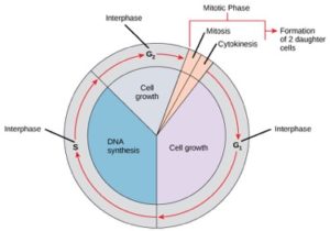
Cells on the path to cell division proceed through a series of precisely timed and carefully regulated stages of growth, DNA replication, and division that produce two genetically identical cells. The cell cycle has two major phases: interphase and the mitotic phase (Figure 14.6.1). During interphase, the cell grows, and DNA is replicated. During the mitotic phase, the replicated DNA and cytoplasmic contents are separated and the cell divides.
Interphase
During interphase, the cell undergoes normal processes while also preparing for cell division. For a cell to move from interphase to the mitotic phase, many internal and external conditions must be met. The three stages of interphase are called G1, S, and G2.
G1 Phase (First Gap)
The first stage of interphase is called the G1 phase (first gap) because, from a microscopic aspect, little change is visible. However, during the G1 stage, the cell is quite active at the biochemical level. The cell is accumulating the building blocks of chromosomal DNA and the associated proteins as well as accumulating sufficient energy reserves to complete the task of replicating each chromosome in the nucleus.
S Phase (Synthesis of DNA)
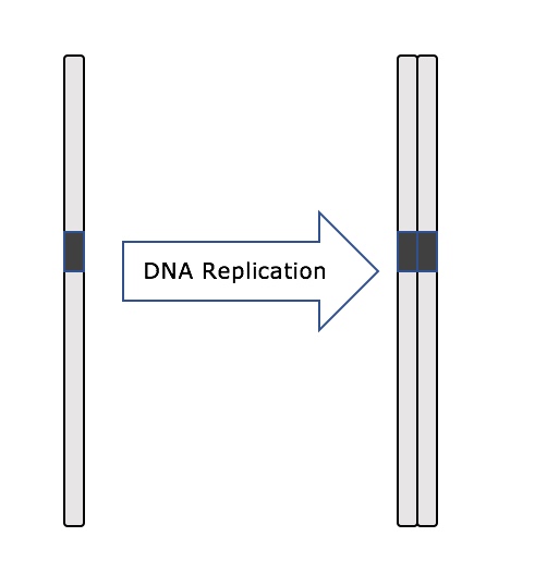
Throughout interphase, nuclear DNA remains in a semi-condensed chromatin configuration. In the S phase, DNA replication can proceed through the mechanisms that result in the formation of identical pairs of DNA molecules—sister chromatids—that are firmly attached to the centromeric region (Figure 14.6.2).
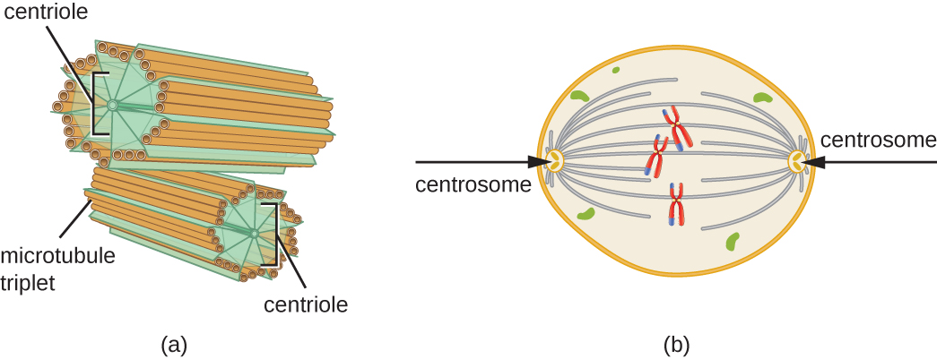
The centrosome is also duplicated during the S phase. The two centrosomes will give rise to the mitotic spindle, the apparatus that orchestrates the movement of chromosomes during mitosis. At the center of each animal cell, the centrosomes of animal cells are associated with a pair of rod-like objects, the centrioles, which are at right angles to each other. Centrioles help organize cell division. Centrioles are not present in the centrosomes of other eukaryotic species, such as plants and most fungi.
G2 Phase (Second Gap)
In the G2 phase, the cell replenishes its energy stores and synthesizes proteins necessary for chromosome manipulation. Some cell organelles are duplicated, and the cytoskeleton is dismantled to provide resources for the mitotic phase. There may be additional cell growth during G2. The final preparations for the mitotic phase must be completed before the cell is able to enter the first stage of mitosis.
The Mitotic Phase
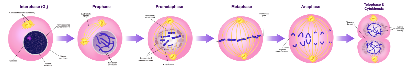
To make two daughter cells, the contents of the nucleus and the cytoplasm must be divided. The mitotic phase is a multistep process during which the duplicated chromosomes are aligned, separated, and moved to opposite poles of the cell, and then the cell is divided into two new identical daughter cells. The first portion of the mitotic phase, mitosis, is composed of five stages, which accomplish nuclear division (Figure 14.6.4). The second portion of the mitotic phase, called cytokinesis, is the physical separation of the cytoplasmic components into two daughter cells. Although the stages of mitosis are similar for most eukaryotes, the process of cytokinesis is quite different for eukaryotes that have cell walls, such as plant cells.
For a review of the details of mitosis, watch the following video from the Amoeba sisters.
Video 14.6.1. The Amazing Cell Process that Uses Division to Multiply! (Updated) by Amoeba Sisters
G0 Phase
Not all cells adhere to the classic cell-cycle pattern in which a newly formed daughter cell immediately enters interphase, closely followed by the mitotic phase. Cells in the G0 phase are not actively preparing to divide. The cell is in a quiescent (inactive) stage, having exited the cell cycle. Some cells enter G0 temporarily until an external signal triggers the onset of G1. Other cells that never or rarely divide, such as mature cardiac muscle and nerve cells, remain in G0 permanently).
Practice Questions
Section Summary
The cell cycle is an orderly sequence of events. Cells on the path to cell division proceed through a series of precisely timed and carefully regulated stages. In eukaryotes, the cell cycle consists of a long preparatory period, called interphase. Interphase is divided into G1, S, and G2 phases. The mitotic phase begins with karyokinesis (mitosis), which consists of five stages. The final stage of the mitotic phase is cytokinesis, during which the cytoplasmic components of the daughter cells are separated either by an actin ring (animal cells) or by cell plate formation (plant cells).
Glossary
- cell cycle
- ordered series of events involving cell growth and cell division that produces two new daughter cells
- centriole
- rod-like structure constructed of microtubules at the center of each animal cell centrosome
- cytokinesis
- division of the cytoplasm following mitosis that forms two daughter cells.
- G0 phase
- distinct from the G1 phase of interphase; a cell in G0 is not preparing to divide
- G1 phase
- (also, first gap) first phase of interphase centered on cell growth during mitosis
- G2 phase
- (also, second gap) third phase of interphase during which the cell undergoes final preparations for mitosis
- interphase
- period of the cell cycle leading up to mitosis; includes G1, S, and G2 phases (the interim period between two consecutive cell divisions
- mitosis
- (also, karyokinesis) period of the cell cycle during which the duplicated chromosomes are separated into identical nuclei; includes prophase, prometaphase, metaphase, anaphase, and telophase
- mitotic phase
- period of the cell cycle during which duplicated chromosomes are distributed into two nuclei and cytoplasmic contents are divided; includes karyokinesis (mitosis) and cytokinesis
- mitotic spindle
- apparatus composed of microtubules that orchestrates the movement of chromosomes during mitosis
- quiescent
- refers to a cell that is performing normal cell functions and has not initiated preparations for cell division
- S phase
- second, or synthesis, stage of interphase during which DNA replication occurs
Figure Descriptions
Figure 14.6.1. The diagram is a circular chart depicting the cell cycle, divided into major phases. Interphase occupies most of the cycle and is split into G1 (cell growth), S (DNA synthesis and centrosome replication), and G2 (further growth and protein synthesis) segments. The smaller mitotic phase section includes mitosis (nuclear division) and cytokinesis (division of the cytoplasm), resulting in the formation of two daughter cells. Red arrows show the cyclical progression through these phases. [Return to Figure 14.6.1]
Figure 14.6.3. Panel (a) shows a close-up view of two cylindrical centrioles positioned perpendicular to each other, each composed of nine sets of microtubule triplets arranged in a ring. Panel (b) illustrates a cell in mitosis with two centrosomes located at opposite poles. From each centrosome, gray threadlike spindle fibers extend toward the center of the cell, connecting to red X-shaped chromosomes aligned in the middle. [Return to Figure 14.6.3]
Figure 14.6.4. A sequential diagram illustrates the process of mitosis. In interphase, the cell contains a nucleus with uncondensed DNA and two centrosomes. During prophase, chromosomes condense, and spindle fibers begin forming from centrosomes at opposite poles. In prometaphase, the nuclear envelope fragments, and spindle fibers attach to chromosomes at their kinetochores. In metaphase, chromosomes align along the cell’s equatorial plane. During anaphase, sister chromatids are pulled apart toward opposite poles. Finally, in telophase and cytokinesis, nuclear envelopes reform around separated chromosomes, and the cell physically divides into two daughter cells. [Return to Figure 14.6.4]
Licenses and Attributions
This chapter, “The Eukaryotic Cell Cycle,” by Elizabeth Dahlhoff, is adapted from “The Cell Cycle” in Biology by Open Stax College (University of Hawaii) under a CC BY-NC 4.0 license. This work is licensed under a CC BY-NC 4.0 license.
Media Attributions
- OSC_Microbio_03_04_Centrosome
- Mitosis_Stages
