21.1 Introduction to Hormones Involved in Glucose Homeostasis
Learning Objectives
After reading this section, you will be able to do the following:
- Describe four types of signaling found in multicellular organisms.
- Describe the function of hormones in multicellular organisms.
- Apply the principles of homeostasis to blood glucose metabolism.
As we learned previously, glucose is a key molecule to drive the growth, maintenance, and reproduction of organisms. Heterotrophs (consumers) must obtain their glucose periodically from feeding on other organisms, which means that the levels of glucose available to cells in multicellular organisms increase dramatically immediately after feeding. During high activity, when muscle cells are contracting at a high frequency, glucose levels in the blood drop, which can be dangerous for the function of other cells such as neurons in the body. However, when glucose blood levels deviate from their normal set point, they are typically very quickly normalized back to those set points between 70 and 100 mg/dl throughout the day. This is a form of homeostasis, a concept that was introduced in Section 11.1, Introduction to Homeostasis. In the video below, you will be introduced to the mechanisms by which this blood glucose homeostasis is achieved.
Video 21.1.1. Blood Glucose Regulation by Janet Lindsley
The ways by which the hormones insulin and glucagon regulate blood glucose levels is a form of endocrine signaling. You should review the introduction to signaling pathways in Section 20.1, Signaling Molecules and Receptors.
Forms of Signaling
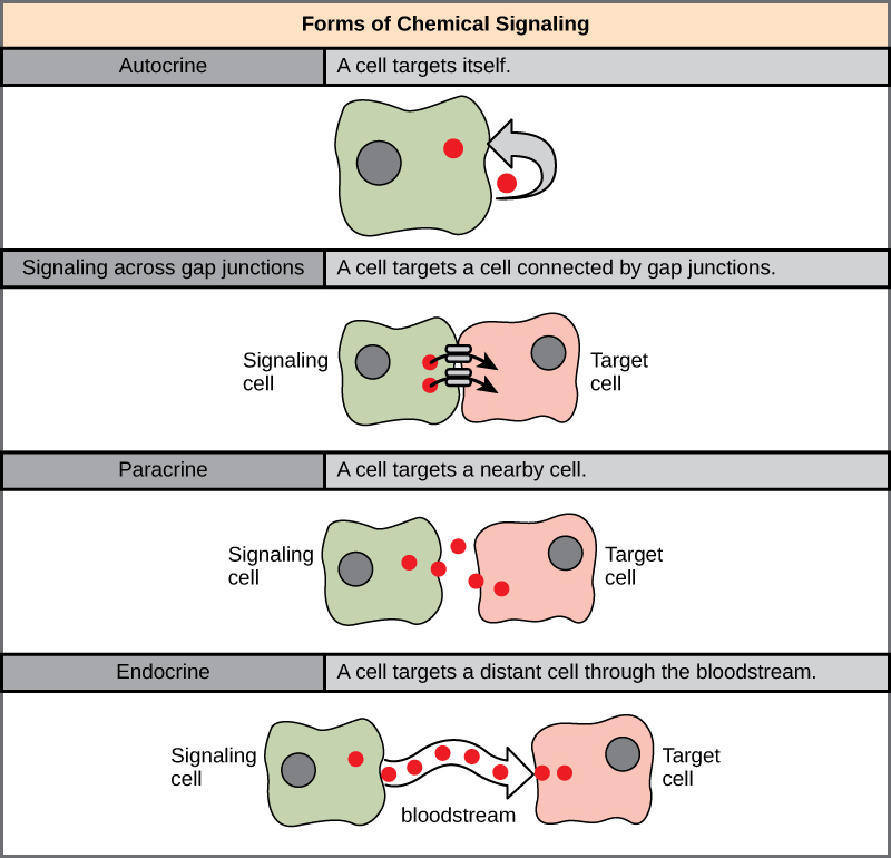
There are four categories of chemical signaling found in multicellular organisms: paracrine signaling, endocrine signaling, autocrine signaling, and direct signaling across gap junctions. The main difference between the different categories of signaling is the distance that the signal travels through the organism to reach the target cell (Figure 21.1.1). Not all cells are affected by the same signals, to cause a response a chemical signal must reach the target cell in high enough concentrations and must bind and trigger activation of its target receptor. Different cell types may respond differently to the signal depending on the type of receptor they express and the downstream signaling response that is initiated.
Blood Glucose Homeostasis Is an Example of Endocrine Signaling
Maintaining homeostasis within the body requires the coordination of many different systems and organs. Communication between neighboring cells, and between cells and tissues in distant parts of the body, occurs through the release of chemicals called hormones. Hormones are released into body fluids (usually blood) that carry these chemicals to their target cells. At the target cells, which are cells that have a receptor for a signal or ligand from a signal cell, the hormones elicit a response. The cells, tissues, and organs that secrete hormones make up the endocrine system. Examples of glands of the endocrine system include the adrenal glands, which produce hormones such as epinephrine and norepinephrine that regulate responses to stress, and the thyroid gland, which produces thyroid hormones that regulate metabolic rates.
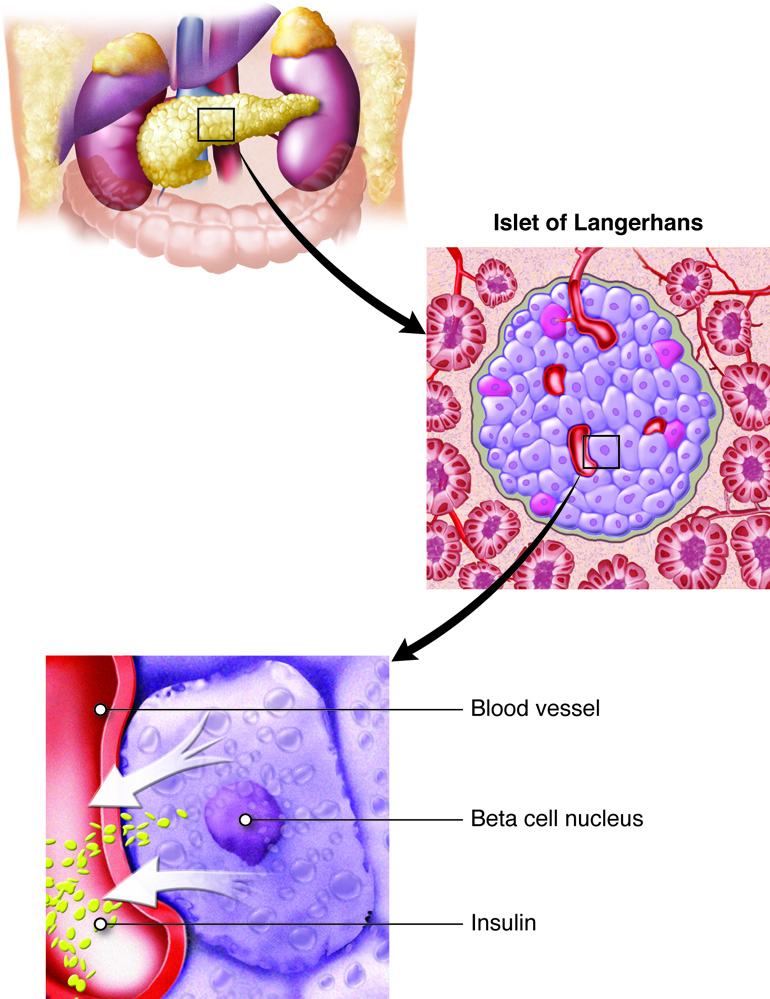
Although there are many different hormones in the human body, they can be divided into three classes based on their chemical structure: lipid-derived, amino acid-derived, and peptide (peptide and proteins) hormones. The two key hormones that regulate blood glucose levels are both peptide hormones and both are produced by specialized endocrine cells in the pancreas (Figure 21.1.2).
Insulin
Insulin is produced by beta cells in the pancreas in response to high blood glucose levels in the bloodstream. Insulin is a initially produced as a 110 amino acid polypeptide called preproinsulin, which then undergoes post-translational modification and protease cleavage to form the final product made up of two amino acid chains held together by disulfide bonds.
Glucagon
Glucagon is produced by alpha cells in the pancreas in response to low blood glucose levels in the bloodstream. Glucagon is a 29 amino acid peptide hormone.
Practice Questions
Glossary
Hormones
Chemicals that ensure the communication between neighboring cells, or between cells in specific tissues with more distant parts of the body.
Autocrine signaling
A cell communicates with itself.
Signaling across gap junctions
A cell communicates via intracellular or membrane signals with a neighboring cell connected via gap junctions (cytoplasmic bridges).
Paracrine signaling
A cell releases a chemical into the extracellular space to communicate with a neighboring cell.
Endocrine signaling
A cell releases a chemical to communicate with distant cells via the bloodstream.
insulin
Peptide hormone released by beta cells of the pancreas in response to high glucose levels in the bloodstream.
glucagon
Peptide hormone released by alpha cells of the pancreas in response to low glucose levels in the bloodstream.
beta cells of the pancreas
Endocrine cells that produce and secrete the hormone insulin.
alpha cells of the pancreas
Endocrine cells that produce and secrete the hormone glucagon.
Figure Descriptions
Figure 21.1.1 The image represents different forms of chemical signaling between cells. It is divided into four sections, each depicting a distinct signaling mode. 1. Autocrine: Shows a single green cell with a gray nucleus releasing a red signal that loops back to itself, indicating self-targeting. 2. Signaling across gap junctions: Illustrates two adjacent cells, one green and one pink. A red signal travels directly between the cells through gap junctions, showing direct communication. 3. Paracrine: Displays a green signaling cell and a pink target cell in close proximity. Red signals move directly to the target cell, indicating nearby cell communication. 4. Endocrine: Shows a green signaling cell releasing red signals into a wavy red bloodstream, targeting a distant pink cell, symbolizing long-distance signaling through the bloodstream. [Return to Figure 21.1.1]
Figure 21.1.2. The image is a detailed illustration showing the structure and function of the pancreas and its cellular components in relation to insulin production. At the top, there is a cross-section of the human pancreas, depicted in yellow and cream colors, against other abdominal organs. A square highlights a specific area, which an arrow points to and magnifies as the Islet of Langerhans. This section features clusters of purple-colored cells surrounded by red blood vessels. Further magnified, another arrow points to an individual beta cell within the islet. This close-up displays a labeled blood vessel in red, beta cell nucleus marked in purple, and insulin depicted as small green dots exiting the cell towards the blood vessel. [Return to Figure 21.1.2]
Licenses and Attributions
“Introduction to Hormones Involved in Glucose Homeostasis” by Christelle Sabatier is adapted from “9.1 Signaling Molecules and Cellular Receptors” by Mary Ann Clark, Matthew Douglas, and Jung Choi for OpenStax Biology 2e used under CC BY 4.0 and from “52 Endocrine Structures and Functions” by SBCCOE for Anatomy & Physiology used under CC BY-NC-SA 4.0. “Introduction to Hormones Involved in Glucose Homeostasis” is licensed under CC BY-NC 4.0.
Media Attributions
- 1C_21_Forms_of_chemical_signaling © OpenStax Biology 2e is licensed under a CC BY (Attribution) license
- 1C_21_Detailed View of Pancreas © CCCOnline is licensed under a CC BY-NC-SA (Attribution NonCommercial ShareAlike) license
