Chapter 13. The Urinary System
13.6 Physiology of Urine Formation: The Vertical Osmotic Gradient and Altering Urine Concentration
Learning Objectives
- Explain the role of the loop of Henle as a countercurrent multiplier in establishing the vertical osmotic gradient in the kidney interstitial fluid
- Explain the role of the vasa recta as a countercurrent exchanger in maintaining the vertical osmotic gradient in the kidney interstitial fluid
- State the two main substances found in the medullary interstitial fluid that contribute to the vertical osmotic gradient
- Describe the effect of ADH on final urine concentration by explaining ADH effects on the distal nephron
- Describe the importance of the vertical osmotic gradient to the kidneys’ ability to excrete concentrated urine
As described earlier, filtrate is the fluid that results from filtration of the plasma into the glomerular capsule. Urine is the waste product resulting from the processing of filtrate: once the filtrate has passed through the entirety of the nephron and exits the collecting ducts, it is called urine. Normally, the kidneys excrete an average of 800 to 2,000 mL of urine per day, but the volume of urine excreted varies depending on several factors. For example, the food and liquids ingested by a person and their hydration status affect urine output. Additionally, certain medications alter the volume of urine produced.
As mentioned in Section 13.0, one of the main functions of the kidneys is osmoregulation, which is maintaining the osmolarity (solute concentration) of the blood. The kidneys maintain plasma solute concentration by varying urine concentration and volume. The concentration of urine produced varies throughout a day depending on the body’s needs. To understand this, consider these extremes:
- when a person is dehydrated and water needs to be conserved, the kidneys produce a small volume of concentrated urine; and
- when a person is overhydrated and excess water needs to be excreted, the kidneys produce a large volume of dilute urine.
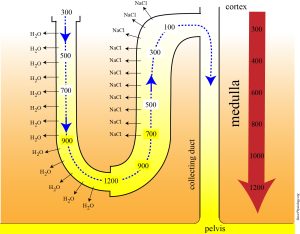
The kidneys’ ability to vary urine concentration and volume relies on the vertical osmotic gradient in the interstitial fluid of the kidney (Figure 13.6.1.)
Countercurrent Mechanisms and Establishment of the Vertical Osmotic Gradient
The vertical osmotic gradient of the interstitial fluid of the kidney is created and maintained countercurrent mechanisms occurring in the Loops of Henle and the vasa recta of juxtamedullary nephrons. The term “countercurrent” refers to opposite fluid flow in adjacent tubes. Two countercurrent systems function in the nephron:
- the countercurrent multiplier system of the Loops of Henle of juxtamedullary nephrons; and
- the countercurrent exchanger system of the vasa recta surrounding the Loops of Henle.
Countercurrent Multiplier System: Establishment of the Vertical Gradient
The longer loops of Henle of juxtamedullary nephrons are countercurrent multipliers. “Countercurrent” refers to the opposite flow of filtrate in the descending and ascending limbs (Figure 13.6.1). “Multiplier” refers to the fact that the osmolarity of the interstitial fluid is multiplied, or increased, as we move from the cortex into the medulla (Figure 13.6.1). This multiplication results from the countercurrent flow of filtrate and the different permeabilities of the descending and ascending limbs. Remember that the descending limb is permeable to water, but not salts, and the ascending limb is permeable to salts, but not water.
The ascending limb actively pumps out salts, mainly NaCl, which adds to the osmolarity of the interstitial fluid. The countercurrent flow in and the different properties of the descending and ascending limbs establish a positive feedback cycle by which this pumping action of the ascending limb is multiplied. The cycle can be summarized as follows:
- the ascending limb pumps NaCl out of the filtrate, which increases the interstitial fluid osmolarity
- the increased interstitial fluid osmolarity causes water to move out of the descending limb via osmosis
- as water leaves the filtrate in the descending limb, its concentration increases, providing salt to be pumped out of the ascending limb (back to step 1)
External Website
Watch this video to help you visualize the role of the loop of Henle as countercurrent multiplier.
Video 13.6.1. Loop of Henle Explained by Biomed Sessions
Urea Recycling and the Vertical Osmotic Gradient
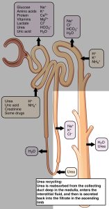
The countercurrent multiplication carried out by the loop of Henle adds NaCl to the interstitial fluid, increasing its osmolarity. NaCl is not the only solute causing this increased osmolarity, however. Urea is also present in the interstitial fluid and contributes to the vertical osmotic gradient. This can be confusing, since urea is a metabolic waste product. Indeed, about 40% to 50% of the urea in the filtrate is eventually excreted in the urine. However, the presence of urea in the interstitial fluid is vital to increasing the osmolarity of the interstitial fluid.
The handling of urea by the nephron is complicated, but here we focus on how urea is added to the interstitial fluid. This is accomplished via the “recycling” of urea between the collecting duct filtrate, the interstitial fluid of the medulla, and the filtrate in the ascending limb (Figure 13.6.2).
Countercurrent Exchanger System: Maintenance of the Vertical Gradient
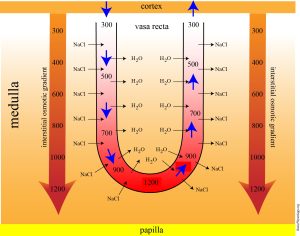
The vasa recta surrounding the loop of Henle functions as a countercurrent exchanger: the flow of blood in the two limbs of the vasa recta is countercurrent and as seen in Figure 13.6.3, the blood flowing through the vasa recta equilibrates with surrounding interstitial fluid. This countercurrent arrangement of flow is important in preventing rapid removal of NaCl from the interstitial fluid and in reabsorbing water. In general, blood flows slowly in capillaries to allow time for exchange of nutrients and wastes. In the vasa recta particularly, this rate of flow is important for two additional reasons. The flow must be slow to allow blood cells to lose and regain water without either crenating or bursting. Second, a rapid flow would remove too much Na+ and urea, destroying the osmotic gradient. As the vasa recta descends, Na+ and urea are freely able to enter the capillary, while water freely leaves; as they ascend, Na+ and urea leave the filtrate into the surrounding medulla, while water reenters the blood.
Formation of Concentrated or Dilute Urine
Section Review
The kidneys maintain plasma solute concentration by varying urine concentration and volume. Their ability to excrete concentrated urine depends on the presence of the vertical osmotic gradient in the interstitial fluid of the kidney. The vertical osmotic gradient is established by the countercurrent multiplier mechanism of the loops of Henle of juxtamedullary nephrons: the ascending limb pumps NaCl into the medullary interstitial fluid, which contributes to the increased osmolarity here. Urea also accumulates in the interstitium due to urea recycling. The vasa recta is a countercurrent exchanger that maintains the vertical osmotic gradient.
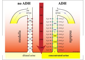
ADH is a signal that water needs to be conserved: as ADH levels rise in response to various stimuli, the permeabilities of the DCT and collecting duct to water increases and water is reabsorbed. This results in the excretion of a smaller volume of concentrated urine. When ADH levels are low, a larger volume of dilute urine is excreted.
Review Questions
Critical Thinking Questions
Glossary
- countercurrent exchange
- mechanism by which vasa recta maintains the vertical osmotic gradient; involves the vasa recta equilibrating with the surrounding interstitial fluid
- countercurrent multiplication
- mechanism by which the ascending and descending limbs of the loop of Henle establish the vertical osmotic gradient; involves pumping of NaCl into the interstitial fluid
- filtrate
- fluid produced by filtration of plasma into the glomerular capsule
- osmoregulation
- maintaining the solute concentration of the blood
- urine
- waste product formed from processing of the filtrate by the nephron; transported from the kidneys to the bladder via the ureters
Glossary Flashcards
This work, Human Physiology, is adapted from Anatomy & Physiology by OpenStax, licensed under CC BY. This edition, with revised content and artwork, is licensed under CC BY-SA except where otherwise noted.
Images from Anatomy & Physiology by OpenStax are licensed under CC BY except where otherwise noted.
Access the original for free at OpenStax.
Image Descriptions
Figure 13.6.1. This diagram illustrates the countercurrent multiplier mechanism in the loop of Henle, showing how the kidney generates and maintains a medullary osmotic gradient. The image depicts the descending and ascending limbs of the loop of Henle, with the cortex at the top (shown in red) and the pelvis at the bottom (shown in yellow). Osmolarity values are labeled along both limbs, starting at 300 mOsm at the cortex and progressively increasing to 1200 mOsm at the bottom of the loop near the pelvis, then decreasing back to 100 mOsm as fluid ascends. The descending limb (left side) is permeable to water (H2O) as indicated by outward arrows, causing the tubular fluid to become progressively more concentrated as it descends. The ascending limb (right side) shows active transport of sodium and chloride (Na+ and Cl-) out of the tubule, as indicated by arrows and symbols, while being impermeable to water. Blue arrows within the tubule indicate the direction of fluid flow down the descending limb and up the ascending limb. A vertical osmolarity scale on the right shows the gradient from cortex to medulla, with a large red arrow indicating increasing osmolarity toward the pelvis. The interstitial fluid osmolarity values (300, 400, 600, 800, 1000, 1200 mOsm) are shown in the space between the limbs, demonstrating the hyperosmotic medullary interstitium. This countercurrent multiplication system enables the kidney to produce concentrated urine by establishing and maintaining the osmotic gradient necessary for water reabsorption in the collecting duct. [Return to Figure 13.6.1]
Figure 13.6.2. This diagram illustrates the ionic concentrations and urea recycling mechanism in the loop of Henle and collecting duct. The image shows a simplified nephron structure with the loop of Henle on the left and the collecting duct on the right (shown in pale orange/tan). At the top, a gray box lists the normal plasma concentrations: Na+ 142 mEq/L, K+ 5 mEq/L, Urea 5 mM, Glucose 5 mM, and Cl- 108 mEq/L. Another box shows ADH (antidiuretic hormone) presence with +/- indicators. Throughout the tubule segments, black boxes with white text indicate the concentrations of Na+, K+, and Cl- at different points, showing how these values change as fluid moves through the system. The descending limb shows Na+ 285, K+ 10, and Cl- 285, while the ascending limb shows Na+ 35, K+ 75, and Cl- 60. At the bottom of the loop, concentrations peak with Na+ and Cl- at 285 mEq/L. Orange arrows indicate the movement of urea, demonstrating urea recycling between the collecting duct and the loop of Henle. A text box at the bottom explains: “Urea recycling: Water reabsorption from the collecting duct concentrates the urea, some of which diffuses back into the filtrate in the ascending loop of Henle.” Additional labels show “ADH +” near the collecting duct, indicating where ADH acts to increase water reabsorption, and “1200 mOsm” indicating the maximum osmolarity achieved at the tip of the loop. [Return to Figure 13.6.2]
Figure 13.6.3. This diagram illustrates the countercurrent multiplier system in the loop of Henle, showing the movement of sodium (Na+) and water (H2O) between the descending and ascending limbs. The image depicts a U-shaped tubule with the cortex at the top (shown in orange) and the papilla at the bottom (shown in yellow). Osmolarity values are labeled at different levels: 300 mOsm in the cortex, progressing to 600 mOsm in the outer medulla, and reaching 1200 mOsm at the deepest point near the papilla. The vasa recta (blood vessels) are shown on both sides of the loop. Blue arrows indicate water (H2O) movement out of the descending limb, while pink/purple arrows show active sodium (Na+) transport out of the ascending limb. The descending limb is permeable to water but relatively impermeable to solutes, allowing water to move into the increasingly concentrated medullary interstitium. The ascending limb actively pumps Na+ and Cl- into the interstitium while being impermeable to water. Large red arrows on the outer edges point downward toward the medulla on the left and upward from the medulla on the right, representing the direction of blood flow in the vasa recta and the maintenance of the osmotic gradient. The color gradient from orange (cortex) through red (medulla) to yellow (papilla) visually represents the increasing osmolarity from the cortex to the inner medulla, demonstrating how this countercurrent system concentrates urine. [Return to Figure 13.6.3]
Figure 13.6.4. This diagram compares the function of the collecting duct in the presence and absence of antidiuretic hormone (ADH). The image is split into two panels showing the collecting duct extending from the medulla (top, shown in orange/red gradient) to the pelvis (bottom, shown in yellow). A large red arrow labeled “ADH” separates the two conditions. The left panel labeled “no ADH” shows the collecting duct with osmolarity measurements ranging from 100 mOsm at the top to 1200 mOsm at the medullary depth, but the tubular fluid remains at approximately 100 mOsm throughout its length, indicated by the pale yellow color within the duct. Small amounts of H2O (water) movement are shown with minimal arrows. This results in dilute urine production. The right panel labeled “ADH” shows the same collecting duct structure, but with ADH present, the tubular fluid osmolarity increases progressively from 300 mOsm at the top to 1200 mOsm at the bottom, matching the surrounding medullary interstitium osmolarity (shown by the deepening yellow color within the duct). Multiple blue arrows indicate increased H2O reabsorption from the duct into the hyperosmotic medullary interstitium. The interstitial osmolarity values are marked at regular intervals (100, 300, 500, 700, 900, 1100, 1200 mOsm) along both ducts. This demonstrates how ADH increases water permeability of the collecting duct by inserting aquaporin channels, allowing water to be reabsorbed and producing concentrated urine, while the absence of ADH results in an impermeable collecting duct that produces dilute urine. [Return to Figure 13.6.4]
Report an Error
Did you find an error, typo, broken link, or other problem in the text? Please follow this link to the error reporting form to submit an error report to the authors.
