Chapter 17. The Endocrine System
17.3 The Pituitary Gland and Hypothalamus
Learning Objectives
By the end of this section, you will be able to:
- Explain the anatomical and functional relationships of the hypothalamus and the posterior and anterior lobes of the pituitary gland
- Identify the two hormones released from the posterior pituitary, their target cells, and principal actions
- Identify the six hormones produced by the anterior lobe of the pituitary gland, their target cells, their principal actions, and regulation by the hypothalamus
- Summarize the direct and indirect effects of growth hormone
- Explain how growth hormone release is regulated by hypothalamic hormones
The hypothalamus–pituitary complex can be thought of as the “command center” of the endocrine system. This complex secretes several hormones that directly produce responses in target tissues, as well as hormones that regulate the synthesis and secretion of hormones of other glands. In addition, the hypothalamus–pituitary complex coordinates the messages of the endocrine and nervous systems. In many cases stimuli received by the nervous system must pass through the hypothalamus–pituitary complex to release hormones that can initiate a response.
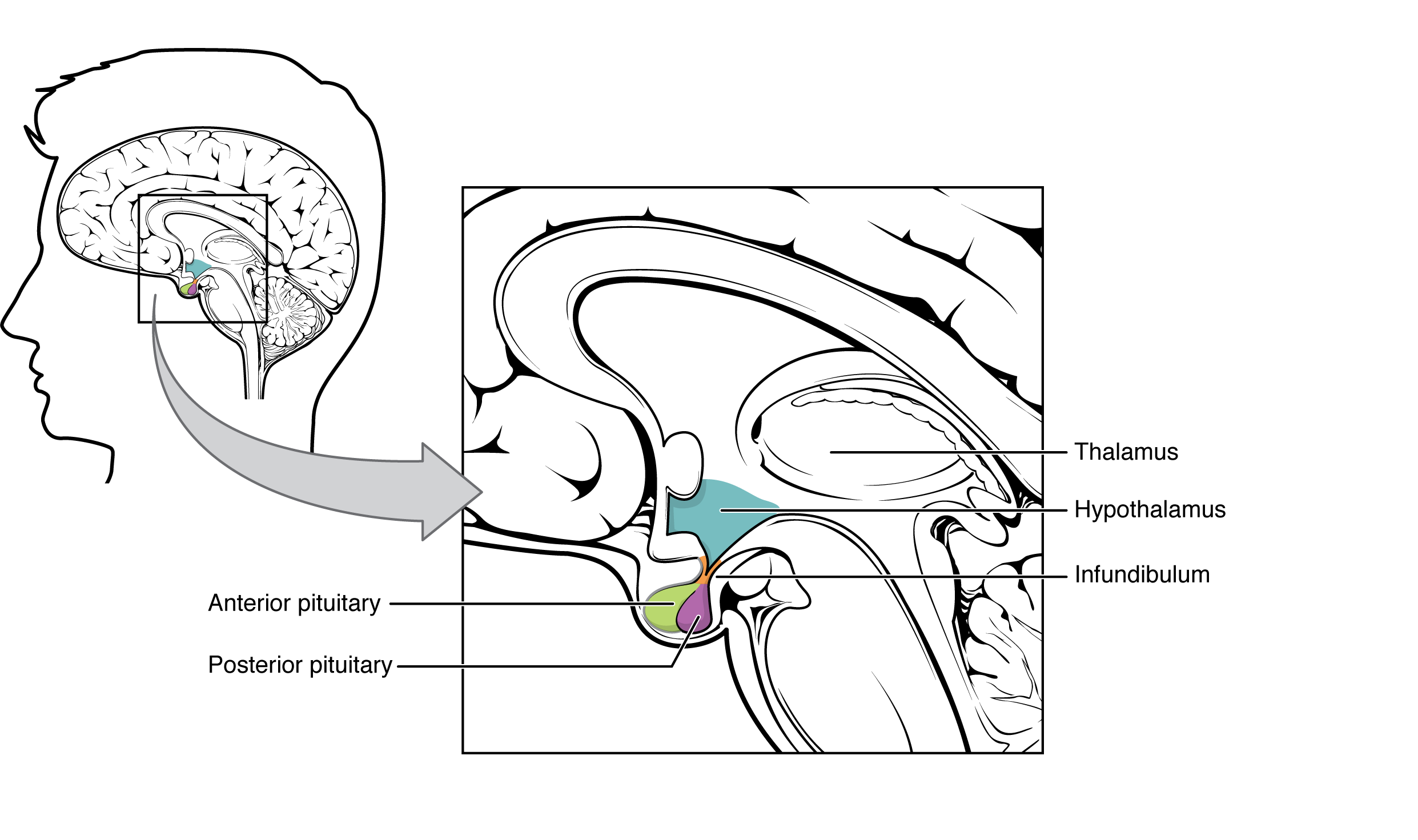
The hypothalamus is a structure of the diencephalon of the brain located anterior and inferior to the thalamus (Figure 17.3.1). It has both neural and endocrine functions, producing and secreting many hormones. In addition, the hypothalamus is anatomically and functionally related to the pituitary gland (or hypophysis), a bean-sized organ suspended from it by a stem called the infundibulum (or pituitary stalk). The pituitary gland consists of two lobes that arise from distinct parts of embryonic tissue: the posterior pituitary (neurohypophysis) is neural tissue, whereas the anterior pituitary (also known as the adenohypophysis [adeno=glandular]) is glandular tissue. The hormones secreted by the posterior and anterior pituitary, and the intermediate zone between the lobes are summarized in Table 17.3.1.
| Pituitary Lobe | Associated Hormones | Chemical Class | Effect |
|---|---|---|---|
| Anterior | Growth hormone (GH) | Protein | Promotes growth of body tissues |
| Anterior | Prolactin (PRL) | Peptide | Promotes milk production from mammary glands |
| Anterior | Thyroid-stimulating hormone (TSH) | Glycoprotein | Stimulates thyroid hormone release from thyroid |
| Anterior | Adrenocorticotropic hormone (ACTH) | Peptide | Stimulates hormone release by adrenal cortex |
| Anterior | Follicle-stimulating hormone (FSH) | Glycoprotein | Stimulates gamete production in gonads |
| Anterior | Luteinizing hormone (LH) | Glycoprotein | Stimulates androgen production by gonads |
| Posterior | Antidiuretic hormone (ADH) | Peptide | Stimulates water reabsorption by kidneys |
| Posterior | Oxytocin | Peptide | Stimulates uterine contractions during childbirth |
| Intermediate zone | Melanocyte-stimulating hormone | Peptide | Stimulates melanin formation in melanocytes |
Posterior Pituitary
The posterior pituitary is actually an extension of the neurons of the paraventricular and supraoptic nuclei of the hypothalamus. The cell bodies of these nuclei are located in the hypothalamus, but their axons descend as the hypothalamic–hypophyseal tract within the infundibulum, and end in axon terminals within the posterior pituitary (Figure 17.3.2).
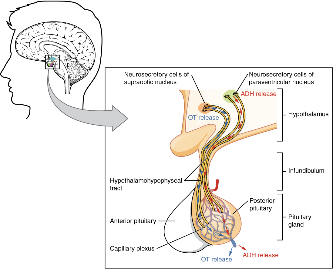
The posterior pituitary gland does not produce hormones, but rather stores and secretes hormones produced by the hypothalamus. Neurons of the paraventricular nucleus produce the hormone oxytocin, whereas neurons of the supraoptic nucleus produce ADH. These hormones travel along the axons into axon terminals within the posterior pituitary. In response to action potentials from the same hypothalamic neurons that produced them, these hormones are released from vesicles within the axon terminals into the bloodstream.
Oxytocin
When fetal development is complete, the peptide-derived hormone oxytocin (“tocia-” meaning “childbirth”) stimulates uterine contractions. Throughout most of pregnancy, oxytocin hormone receptors are not expressed at high levels in the uterus. Toward the end of pregnancy, the synthesis of oxytocin receptors in the uterus increases, and the smooth muscle cells of the uterus become more sensitive to its effects. Oxytocin is continually released throughout childbirth through a positive feedback mechanism. As noted earlier, oxytocin prompts uterine contractions that push the fetal head toward the cervix. In response, cervical stretching stimulates additional oxytocin to be synthesized by the hypothalamus and released from the posterior pituitary. This increases the intensity and effectiveness of uterine contractions and prompts additional stretching of the cervix. This positive feedback loop continues until birth.
Although the mother’s high blood levels of oxytocin begin to decrease immediately following birth, oxytocin continues to play a role in maternal and newborn health. First, oxytocin is necessary for the milk ejection reflex (commonly referred to as “let-down”) in breastfeeding women. As the newborn begins suckling, sensory receptors in the nipples transmit signals to the hypothalamus. In response, oxytocin is secreted and released into the bloodstream. Within seconds, myoepithelial cells around the glandular cells of the mammary glands contract, ejecting milk into the infant’s mouth. Secondly, in both males and females, oxytocin is thought to contribute to parent–newborn bonding, known as attachment. Oxytocin is also thought to be involved in feelings of love and closeness, as well as in the sexual response. In the last examples oxytocin is functioning as a neurotransmitter in the brain.
Antidiuretic Hormone (ADH)
The solute concentration of the blood, or blood osmolarity, may change in response to the consumption of certain foods and fluids, as well as in response to disease, injury, medications, or other factors. Blood osmolarity is constantly monitored by osmoreceptors—specialized cells within the hypothalamus that are particularly sensitive to the concentration of sodium ions and other solutes.
In response to high blood osmolarity, which can occur during dehydration or following a very salty meal, stimulation of osmoreceptors causes neurons in the supraoptic nucleus to signal the posterior pituitary to release antidiuretic hormone (ADH). The target cells of ADH are located in the tubular cells of the kidneys. Its effect is to increase epithelial permeability to water, allowing increased water reabsorption. This increase in water reabsorption has the effect of making the blood more dilute and the urine more concentrated. ADH is also known as vasopressin because in high concentrations, rarely seen except in cases of hemorrhage or shock, it causes constriction of blood vessels, which increases blood pressure by increasing peripheral resistance. The release of ADH is controlled by a negative feedback loop. As blood osmolarity decreases, the hypothalamic osmoreceptors sense the change prompting a corresponding decrease in the secretion of ADH. As a result, less water is reabsorbed from the urine filtrate.
Some drugs can affect the secretion of ADH. For example, alcohol consumption inhibits the release of ADH, resulting in increased urine production that can eventually lead to the dehydration of a hangover. A disease called diabetes insipidus is characterized by chronic underproduction of ADH that causes chronic dehydration. Because little ADH is produced and secreted, not enough water is reabsorbed by the kidneys. Although patients feel thirsty, and increase their fluid consumption, this doesn’t effectively decrease the solute concentration in their blood because ADH levels are not high enough to trigger water reabsorption in the kidneys. Electrolyte imbalances can occur in severe cases of diabetes insipidus. The opposite occurs in a condition called SIADH (syndrome of inappropriate antidiuretic hormone), ADH is secreted at extremely high levels leading to extremely concentrated urine producing a dangerously low blood osmolarity known as water intoxication.
Anterior Pituitary
Recall that the posterior pituitary does not synthesize hormones, but merely stores them. In contrast, the anterior pituitary does manufacture hormones. Like the posterior pituitary the release of hormones from the anterior pituitary is controlled by the hypothalamus. This control is mediated by secretion of releasing or inhibiting hormones into the blood.
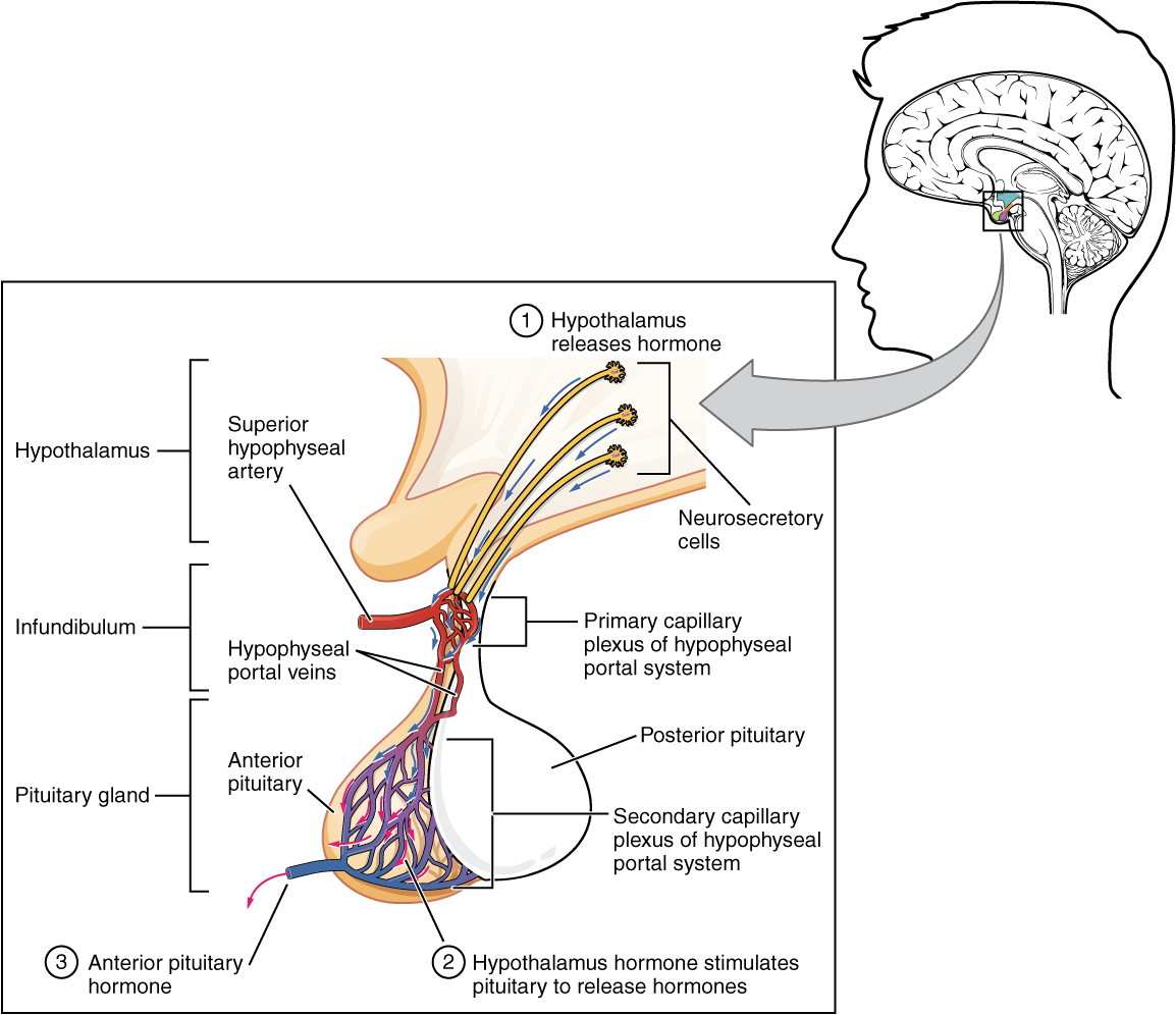
Within the infundibulum is a bridge of capillaries that connects the hypothalamus to the anterior pituitary. This network, called the hypophyseal portal system, allows hypothalamic hormones to be transported to the anterior pituitary without becoming diluted in systemic circulation. This portal system begins with a primary capillary plexus originating from the superior hypophyseal artery, a branches of the internal carotid artery. Blood from the first capillary bed supplies a secondary capillary plexus in the anterior pituitary via the hypophyseal portal veins (Figure 17.3.3). Hypothalamic releasing and inhibiting hormones are released into the primary capillary plexus which drain into the portal veins carrying them to the secondary capillary plexus where they stimulate (or inhibit) the endocrine cells of the anterior pituitary. Hormones produced by the anterior pituitary (in response to hypothalamic releasing hormones) enter the secondary capillary plexus continuing into general circulation.
The anterior pituitary produces seven hormones. These are growth hormone (GH), thyroid-stimulating hormone (TSH), adrenocorticotropic hormone (ACTH), follicle-stimulating hormone (FSH), luteinizing hormone (LH), beta endorphin, and prolactin. Of the hormones of the anterior pituitary, TSH, ACTH, FSH, and LH are collectively referred to as tropic hormones (trope- = “turning”) because they stimulate or inhibit secretion of hormones from other glands. Growth hormone will go on and act on its targets once released from the anterior pituitary.
Growth Hormone
Growth hormone (GH), also called somatotropin regulates the growth of the human body, protein synthesis, and cellular replication. Its primary function is anabolic; it promotes protein synthesis and tissue building through direct and indirect mechanisms (Figure 17.3.4). GH levels are controlled by the release of GHRH and GHIH (also known as somatostatin) from the hypothalamus.
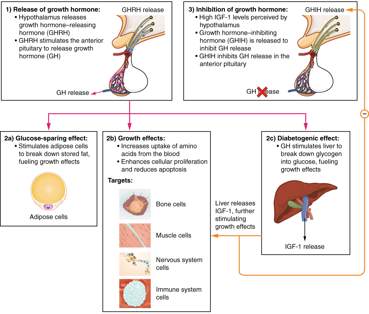
A glucose-sparing effect occurs when GH stimulates lipolysis, or the breakdown of adipose tissue, releasing fatty acids into the blood. As a result, many tissues switch from glucose to fatty acids as their main energy source, which means that less glucose is taken up from the bloodstream.
GH also initiates the diabetogenic effect in which GH stimulates the liver to break down glycogen to glucose, which is then released into the blood. The name “diabetogenic” is derived from the similarity in elevated blood glucose levels observed between individuals with untreated diabetes mellitus and individuals experiencing GH excess. Blood glucose levels rise as the result of a combination of glucose-sparing and diabetogenic effects.
GH indirectly mediates growth and protein synthesis by triggering the liver and other tissues to produce a group of proteins called insulin-like growth factors (IGFs). These proteins enhance cellular proliferation and inhibit apoptosis, or programmed cell death. IGFs stimulate cells to increase their uptake of amino acids from the blood for protein synthesis. Skeletal muscle and cartilage cells are particularly sensitive to stimulation from IGFs.
Dysfunction of the endocrine system’s control of growth can result in several disorders. For example, gigantism is a disorder caused by the hypersecretion of GH before the growth plates have closed resulting in excessive growth of all bones. Hypersecretion of GH after the growth plates have closed results in a condition called acromegaly, a disorder that results in the growth of bones in the face, hands, and feet. Abnormally low levels of GH in children can cause growth impairment—a disorder called pituitary dwarfism (also known as growth hormone deficiency) which affects all bones. Achondroplastic dwarfism affects only the bones with growth plates (long bones) resulting in short arms and legs with normal sized trunk and head.
Thyroid-Stimulating Hormone
The activity of the thyroid gland is regulated by thyroid-stimulating hormone (TSH), also called thyrotropin. TSH is released from the anterior pituitary in response to thyrotropin-releasing hormone (TRH) from the hypothalamus. As will be discussed shortly, it triggers the secretion of thyroid hormones by the thyroid gland. In a classic negative feedback loop, elevated levels of thyroid hormones in the bloodstream then trigger a drop in production of TRH and TSH.
Adrenocorticotropic Hormone
The adrenocorticotropic hormone (ACTH), also called corticotropin, stimulates the adrenal cortex (the more superficial “bark” of the adrenal glands) to secrete corticosteroid hormones such as cortisol. ACTH come from a precursor molecule known as pro-opiomelanotropin (POMC) which produces several biologically active molecules when cleaved, including ACTH, melanocyte-stimulating hormone, and the brain opioid peptides known as endorphins.
The release of ACTH is regulated by the corticotropin-releasing hormone (CRH) from the hypothalamus in response to normal physiologic rhythms. A variety of stressors can also influence its release, and the role of ACTH in the stress response is discussed later in this chapter.
Follicle-Stimulating Hormone and Luteinizing Hormone
The endocrine glands secrete a variety of hormones that control the development and regulation of the reproductive system (these glands include the anterior pituitary, the adrenal cortex, and the gonads—the testes in males and the ovaries in females). Much of the development of the reproductive system occurs during puberty and is marked by the development of sex-specific characteristics in both male and female adolescents. Puberty is initiated by gonadotropin-releasing hormone (GnRH), a hormone produced and secreted by the hypothalamus. GnRH stimulates the anterior pituitary to secrete gonadotropins—hormones that regulate the function of the gonads. The levels of GnRH are regulated through a negative feedback loop; high levels of reproductive hormones inhibit the release of GnRH. Throughout life, gonadotropins regulate reproductive function.
The gonadotropins include two glycoprotein hormones: follicle-stimulating hormone (FSH) stimulates the production and maturation of sex cells, or gametes, including ova in women and sperm in men. FSH also promotes ovarian follicular growth in women; these follicles then release estrogens. Luteinizing hormone (LH) triggers ovulation in women, as well as the production of estrogens and progesterone by the ovaries. LH stimulates production of testosterone by the male testes.
Prolactin
As its name implies, prolactin (PRL) promotes lactation (milk production) in women. After birth, it stimulates the mammary glands to produce breast milk. However, the effects of prolactin depend heavily upon the permissive effects of estrogens, progesterone, and other hormones. And as noted earlier, the let-down of milk occurs in response to stimulation from oxytocin.
In a non-pregnant woman, prolactin secretion is inhibited by prolactin-inhibiting hormone (PIH), which is actually the neurotransmitter dopamine, released from neurons in the hypothalamus. Only during pregnancy do prolactin levels rise primarily in response to a decrease in inhibition by PIH and partially due to stimulation by prolactin-releasing hormone (PRH) from the hypothalamus.
Intermediate Pituitary: Melanocyte-Stimulating Hormone
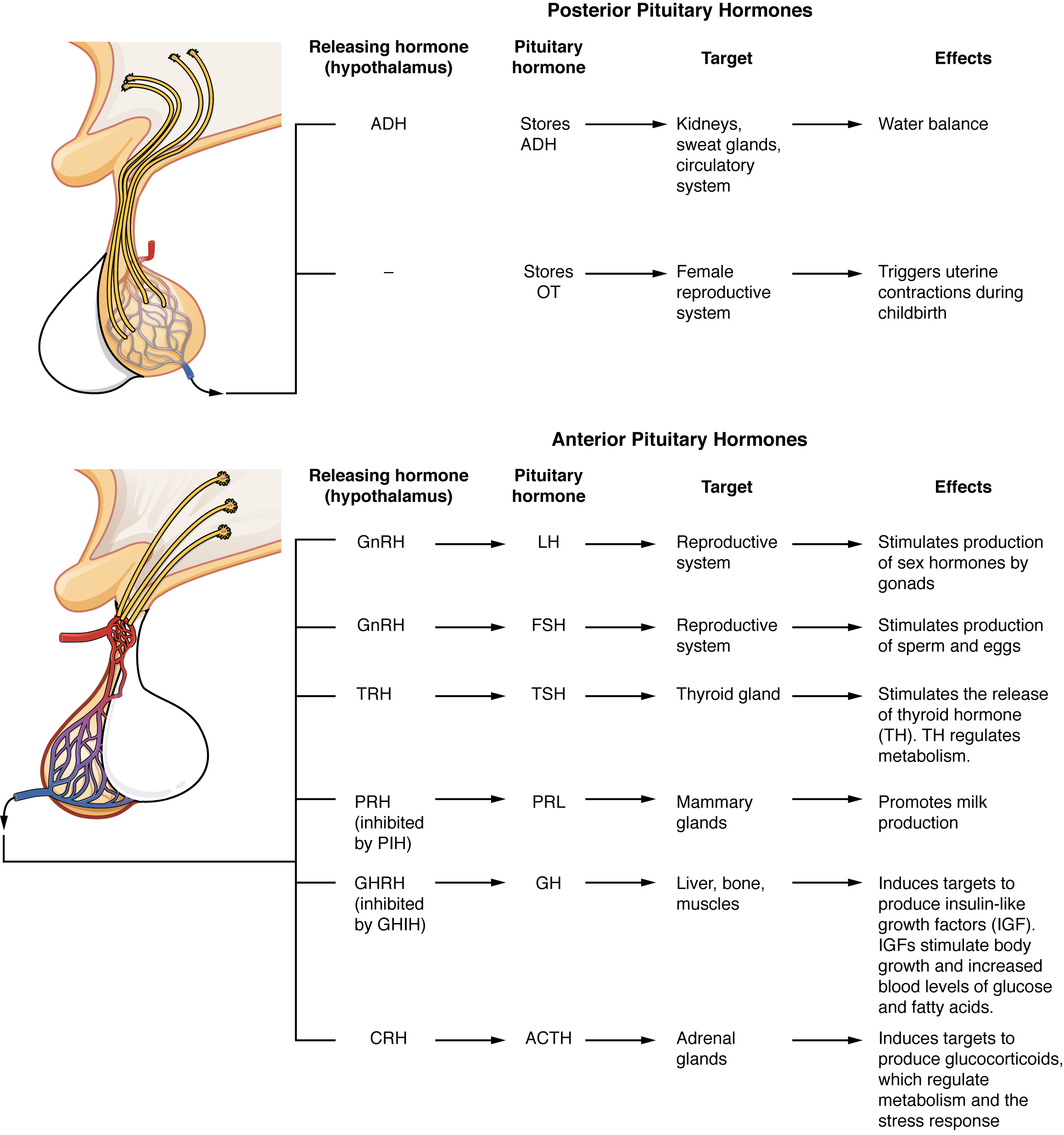
The cells in the zone between the pituitary lobes secrete a hormone known as melanocyte-stimulating hormone (MSH) that is formed by cleavage of the pro-opiomelanocortin (POMC) precursor protein. Local production of MSH in the skin is responsible for melanin production in response to UV light exposure. The role of MSH made by the pituitary is more complicated. For instance, people with lighter skin generally have the same amount of MSH as people with darker skin. Nevertheless, this hormone is capable of darkening of the skin by inducing melanin production in the skin’s melanocytes. Women also show increased MSH production during pregnancy; in combination with estrogens, it can lead to darker skin pigmentation, especially the skin of the areolas and labia minora. Figure 17.3.5 is a summary of the pituitary hormones and their principal effects.
External Website

Visit this link [New Tab] to watch an animation showing the role of the hypothalamus and the pituitary gland.
Section Review
The hypothalamus–pituitary complex is located in the diencephalon of the brain. The hypothalamus and the pituitary gland are connected by a structure called the infundibulum, which contains vasculature and nerve axons. The pituitary gland is divided into two distinct structures with different embryonic origins. The posterior lobe houses the axon terminals of hypothalamic neurons. It stores and releases into the bloodstream two hypothalamic hormones: oxytocin and antidiuretic hormone (ADH). The anterior lobe is connected to the hypothalamus by vasculature in the infundibulum and produces and secretes six hormones. Their secretion is regulated, however, by releasing and inhibiting hormones from the hypothalamus. The six anterior pituitary hormones are: growth hormone (GH), thyroid-stimulating hormone (TSH), adrenocorticotropic hormone (ACTH), follicle-stimulating hormone (FSH), luteinizing hormone (LH), and prolactin (PRL).
Review Questions
Critical Thinking Questions
Glossary
- acromegaly
- disorder in adults caused when abnormally high levels of GH trigger growth of bones in the face, hands, and feet
- adrenocorticotropic hormone (ACTH)
- anterior pituitary hormone that stimulates the adrenal cortex to secrete corticosteroid hormones (also called corticotropin)
- antidiuretic hormone (ADH)
- hypothalamic hormone that is stored by the posterior pituitary and that signals the kidneys to reabsorb water
- follicle-stimulating hormone (FSH)
- anterior pituitary hormone that stimulates the production and maturation of sex cells
- gigantism
- disorder in children caused when abnormally high levels of GH prompt excessive growth
- gonadotropins
- hormones that regulate the function of the gonads
- growth hormone (GH)
- anterior pituitary hormone that promotes tissue building and influences nutrient metabolism (also called somatotropin)
- hypophyseal portal system
- network of blood vessels that enables hypothalamic hormones to travel into the anterior lobe of the pituitary without entering the systemic circulation
- hypothalamus
- region of the diencephalon inferior to the thalamus that functions in neural and endocrine signaling
- insulin-like growth factors (IGF)
- protein that enhances cellular proliferation, inhibits apoptosis, and stimulates the cellular uptake of amino acids for protein synthesis
- luteinizing hormone (LH)
- anterior pituitary hormone that triggers ovulation and the production of ovarian hormones in females, and the production of testosterone in males
- osmoreceptor
- hypothalamic sensory receptor that is stimulated by changes in solute concentration (osmotic pressure) in the blood
- oxytocin
- hypothalamic hormone stored in the posterior pituitary gland and important in stimulating uterine contractions in labor, milk ejection during breastfeeding, and feelings of attachment (also produced in males)
- pituitary dwarfism
- disorder in children caused when abnormally low levels of GH result in growth retardation
- pituitary gland
- bean-sized organ suspended from the hypothalamus that produces, stores, and secretes hormones in response to hypothalamic stimulation (also called hypophysis)
- prolactin (PRL)
- anterior pituitary hormone that promotes development of the mammary glands and the production of breast milk
- thyroid-stimulating hormone (TSH)
- anterior pituitary hormone that triggers secretion of thyroid hormones by the thyroid gland (also called thyrotropin)
Glossary Flashcards
This work, Human Physiology, is adapted from Anatomy & Physiology by OpenStax, licensed under CC BY. This edition, with revised content and artwork, is licensed under CC BY-SA except where otherwise noted.
Images from Anatomy & Physiology by OpenStax are licensed under CC BY except where otherwise noted.
Access the original for free at OpenStax.
Image Descriptions
Figure 17.3.1. This anatomical diagram illustrates the location and relationship of the hypothalamus and pituitary gland within the human brain, shown in a sagittal (side) view. On the left, a profile of a human head shows the brain with a rectangular region highlighted, which is magnified in the detailed inset on the right. The magnified view reveals the hypothalamus (colored in light blue) positioned above the pituitary gland in the central region of the brain, just below the thalamus. The pituitary gland is shown in two distinct parts: the anterior pituitary (colored in light green) and the posterior pituitary (colored in purple). Below the pituitary complex, the infundibulum (also called the pituitary stalk) is labeled, showing the physical connection between the hypothalamus and pituitary gland. An arrow from the smaller brain overview points to the enlarged detail, emphasizing the small but crucial location of these endocrine structures at the base of the brain. This diagram effectively demonstrates the anatomical proximity and connection between the hypothalamus and pituitary gland, which is essential for understanding their coordinated role in regulating the endocrine system through hormone secretion and the hypothalamic-pituitary axis. [Return to Figure 17.3.1]
Figure 17.3.2. This diagram illustrates the detailed structural and functional relationship between the hypothalamus and pituitary gland, showing how hormones are released into the bloodstream. On the left, a sagittal view of a human head shows the brain with a small boxed region indicating the location of the hypothalamic-pituitary complex. The main diagram on the right provides a magnified view of this region, revealing the intricate connection between these structures. The hypothalamus is shown at the top, containing two distinct groups of neurosecretory cells: those in the supraoptic nucleus (shown on the left) that release oxytocin (OT), and those in the paraventricular nucleus (shown on the right) that release antidiuretic hormone (ADH). These neurosecretory cells extend axons down through the infundibulum (pituitary stalk) via the hypothalamohypophyseal tract, depicted as multiple colored nerve fibers. The pituitary gland at the bottom is divided into two lobes: the anterior pituitary and the posterior pituitary. The anterior pituitary contains a dense capillary plexus (shown as an intricate network of blood vessels), while the posterior pituitary receives direct neural connections from the hypothalamus. The diagram shows OT release (marked in blue) occurring both at the top near the hypothalamus and at the bottom near the pituitary, and ADH release (marked in red) similarly occurring at both locations, illustrating how these hormones are transported through axons and released into the bloodstream via the capillary networks. This anatomical arrangement demonstrates the neural and vascular connections that enable the hypothalamus to control endocrine function through direct hormone release in the posterior pituitary and indirect regulation of the anterior pituitary through the portal blood system. [Return to Figure 17.3.2]
Figure 17.3.3. This anatomical diagram illustrates the hypothalamic-pituitary portal system, showing how the hypothalamus regulates anterior pituitary hormone secretion through a specialized vascular connection. In the upper right corner, a sagittal view of a human head shows the brain with a small boxed region indicating the location of the hypothalamic-pituitary complex. The main diagram provides a detailed magnified view of this region, revealing the three-step process of hormonal control. At the top, labeled as Step 1, neurosecretory cells in the hypothalamus release hormones (shown as yellow nerve fibers with dark endings) into the superior hypophyseal artery. These hypothalamic hormones enter the primary capillary plexus of the hypophyseal portal system, a network of blood vessels (shown in red) located in the infundibulum (pituitary stalk). The blood then flows downward through the hypophyseal portal veins into the anterior pituitary, where it forms the secondary capillary plexus of the hypophyseal portal system, depicted as an extensive network of purple blood vessels surrounding the anterior pituitary tissue. Step 2 shows how hypothalamus hormones delivered through this portal system stimulate the anterior pituitary to release its own hormones. At the bottom, Step 3 indicates that anterior pituitary hormones are then secreted into the general bloodstream (shown in blue) to reach target organs throughout the body. The posterior pituitary is shown adjacent to the anterior pituitary but is not directly connected to the portal system, reflecting its different mode of hormone release. This diagram effectively demonstrates the unique hypothalamic-pituitary portal circulation, which allows hypothalamic releasing and inhibiting hormones to reach the anterior pituitary in high concentrations without being diluted in the general circulation, enabling precise endocrine control. [Return to Figure 17.3.3]
Figure 17.3.4. This comprehensive diagram illustrates the regulation and multifaceted effects of growth hormone (GH) through the hypothalamic-pituitary axis, including negative feedback control. The diagram is divided into three main sections. Section 1, labeled “Release of growth hormone,” shows the hypothalamus releasing growth hormone-releasing hormone (GHRH), depicted with neurosecretory cells extending from the hypothalamus through the infundibulum to the pituitary gland. GHRH stimulates the anterior pituitary (shown with an extensive purple capillary network) to release growth hormone (GH) into the bloodstream. Section 3, labeled “Inhibition of growth hormone,” demonstrates the negative feedback mechanism where high levels of insulin-like growth factor-1 (IGF-1) are perceived by the hypothalamus, triggering the release of growth hormone-inhibiting hormone (GHIH, also called somatostatin). GHIH then inhibits GH release in the anterior pituitary, shown with a red X over “GH release” and an orange feedback loop with a minus sign indicating inhibition. The middle and lower sections detail the three major categories of GH effects, connected by pink arrows from the GH release. Section 2a shows the “Glucose-sparing effect,” illustrating how GH stimulates adipose cells (depicted as a large yellow fat cell) to break down stored fat, thereby fueling growth effects while conserving glucose. Section 2b, titled “Growth effects,” explains that GH increases uptake of amino acids from the blood and enhances cellular proliferation while reducing apoptosis. This section identifies four major target tissues with microscopic images: bone cells, muscle cells, nervous system cells (showing neurons with branching processes), and immune system cells. Section 2c presents the “Diabetogenic effect,” showing the liver (depicted in brown) where GH stimulates glycogen breakdown into glucose to fuel growth effects. The liver also releases IGF-1, which further stimulates growth effects in target tissues (indicated by an orange arrow connecting back to the growth effects section). This IGF-1 release completes the regulatory loop by providing the negative feedback signal to the hypothalamus shown in Section 3. Together, this diagram demonstrates how growth hormone serves as a master regulator of growth and metabolism through direct effects on multiple tissues and indirect effects mediated by IGF-1, all controlled by a sophisticated feedback system involving the hypothalamus and pituitary gland. [Return to Figure 17.3.4]
Figure 17.3.5. This comprehensive diagram presents the hormones produced by both the posterior and anterior pituitary glands, organized in two detailed tables with accompanying anatomical illustrations. The top section, titled “Posterior Pituitary Hormones,” shows an anatomical illustration of the hypothalamus connected to the posterior pituitary via neurosecretory cells (depicted as yellow nerve fibers). The table lists two hormones: antidiuretic hormone (ADH), which is released by the hypothalamus and stored in the posterior pituitary as ADH, targets the kidneys, sweat glands, and circulatory system to regulate water balance; and oxytocin (OT), which has no listed hypothalamic releasing hormone, is stored in the posterior pituitary as OT, targets the female reproductive system, and triggers uterine contractions during childbirth. The bottom section, titled “Anterior Pituitary Hormones,” displays an illustration showing the hypothalamus connected to the anterior pituitary through the hypophyseal portal system (depicted with red blood vessels feeding into a purple capillary network within the anterior pituitary). This table lists six hormone pathways: gonadotropin-releasing hormone (GnRH) from the hypothalamus stimulates release of luteinizing hormone (LH), which targets the reproductive system to stimulate sex hormone production by gonads; GnRH also stimulates follicle-stimulating hormone (FSH), targeting the reproductive system to stimulate sperm and egg production; thyrotropin-releasing hormone (TRH) stimulates thyroid-stimulating hormone (TSH), which targets the thyroid gland to stimulate release of thyroid hormone (TH) that regulates metabolism; prolactin-releasing hormone (PRH), which is inhibited by prolactin-inhibiting hormone (PIH), stimulates prolactin (PRL) that targets mammary glands to promote milk production; growth hormone-releasing hormone (GHRH), which is inhibited by growth hormone-inhibiting hormone (GHIH), stimulates growth hormone (GH) that targets the liver, bone, and muscles to induce production of insulin-like growth factors (IGF), which stimulate body growth and increase blood levels of glucose and fatty acids; and corticotropin-releasing hormone (CRH) stimulates adrenocorticotropic hormone (ACTH), which targets the adrenal glands to induce production of glucocorticoids that regulate metabolism and the stress response. Together, these tables provide a complete overview of how the hypothalamus regulates both divisions of the pituitary gland through different mechanisms—direct neural connections for the posterior pituitary and releasing/inhibiting hormones delivered through the portal blood system for the anterior pituitary—to control diverse physiological processes including reproduction, growth, metabolism, stress response, lactation, and fluid balance. [Return to Figure 17.3.5]
Report an Error
Did you find an error, typo, broken link, or other problem in the text? Please follow this link to the error reporting form to submit an error report to the authors.