Chapter 17. The Endocrine System
17.5 The Parathyroid Glands
Learning Objectives
By the end of this section, you will be able to:
- Describe the location and structure of the parathyroid glands
- Indicate the general functions of parathyroid hormone
- Discuss how PTH release is regulated
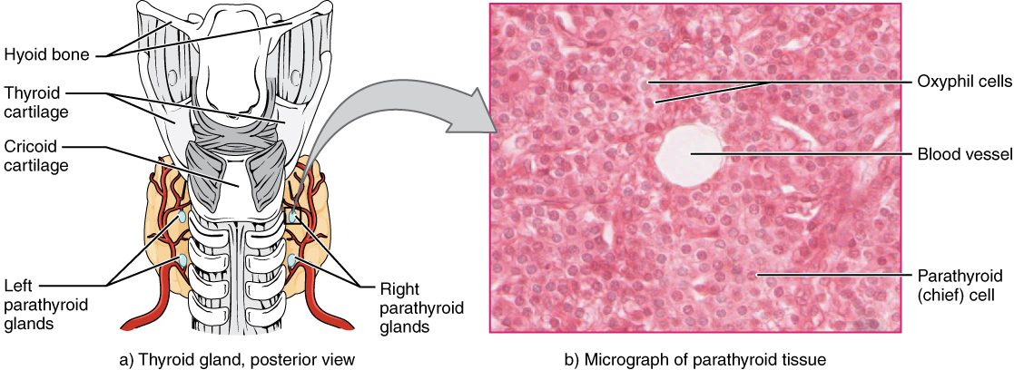
The parathyroid glands are tiny, round structures usually found embedded in the posterior surface of the thyroid gland (Figure 17.5.1). A thick connective tissue capsule separates the glands from the thyroid tissue. Most people have four parathyroid glands, but occasionally there are more in tissues of the neck or chest. The primary functional cells of the parathyroid glands are the chief cells. These epithelial cells produce and secrete the parathyroid hormone (PTH), the major hormone involved in the regulation of blood calcium levels. The gland also contains oxyphil cells but their function is not clear.
Regulation of Release and Functions of PTH
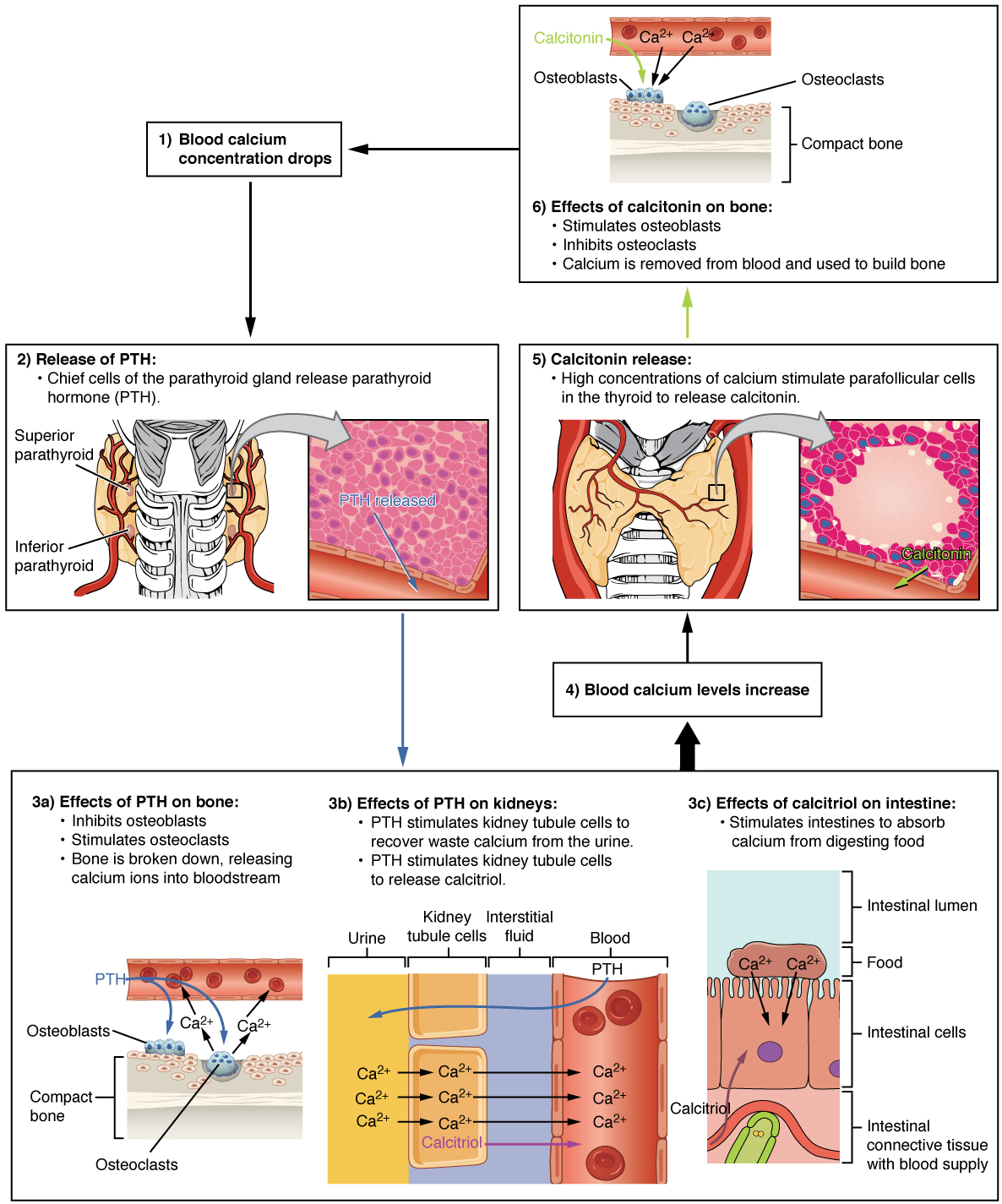
The parathyroid glands produce and secrete PTH, a peptide hormone, in response to low blood calcium levels (Figure 17.5.2). PTH secretion causes the release of calcium from the bones by stimulating osteoclasts, which secrete enzymes that degrade bone and release calcium into the interstitial fluid. PTH also inhibits osteoblasts, the cells involved in bone deposition, thereby sparing blood calcium. PTH causes increased reabsorption of calcium (and magnesium) in the kidney tubules from the urine filtrate. In addition, PTH initiates the production of the steroid hormone calcitriol (also known as 1,25-dihydroxyvitamin D), which is the active form of vitamin D3, in the kidneys. Calcitriol then stimulates increased absorption of dietary calcium by the intestines. This is considered an indirect effect of PTH. A negative feedback loop regulates the levels of PTH, with rising blood calcium levels inhibiting further release of PTH.
Disorders of the Endocrine System – Hyperparathyroidism and Hypoparathyroidism
Abnormally high activity of the parathyroid gland can cause hyperparathyroidism, a disorder caused by an overproduction of PTH that results in excessive calcium reabsorption from bone. Hyperparathyroidism can significantly decrease bone density, leading to spontaneous fractures or deformities. As blood calcium levels rise, cell membrane permeability to sodium is decreased, and the responsiveness of the nervous system is reduced. At the same time, calcium phosphate deposits may collect in the body’s tissues and organs (extraosseous calcification), impairing their functioning.
In contrast, abnormally low blood calcium levels may be caused by parathyroid hormone deficiency, called hypoparathyroidism, which may develop following injury or surgery involving the thyroid gland. Low blood calcium increases membrane permeability to sodium, resulting in muscle twitching, cramping, spasms, or convulsions. Severe deficits can paralyze muscles, including those involved in breathing, and can be fatal.
Section Review
Calcium is required for a variety of important physiologic processes, including neuromuscular functioning; thus, blood calcium levels are closely regulated. The parathyroid glands are small structures located on the posterior thyroid gland that produce parathyroid hormone (PTH), which regulates blood calcium levels. Low blood calcium levels cause the production and secretion of PTH. PTH increases bone resorption, calcium absorption from the intestines, and calcium reabsorption by the kidneys In contrast, elevated blood calcium levels inhibit secretion of PTH and trigger secretion of the thyroid hormone calcitonin. Underproduction of PTH can result in hypoparathyroidism. In contrast, overproduction of PTH can result in hyperparathyroidism.
Review Questions
Critical Thinking Questions
Glossary
- hyperparathyroidism
- disorder caused by overproduction of PTH that results in abnormally elevated blood calcium
- hypoparathyroidism
- disorder caused by underproduction of PTH that results in abnormally low blood calcium
- parathyroid glands
- small, round glands embedded in the posterior thyroid gland that produce parathyroid hormone (PTH)
- parathyroid hormone (PTH)
- peptide hormone produced and secreted by the parathyroid glands in response to low blood calcium levels
Glossary Flashcards
This work, Human Physiology, is adapted from Anatomy & Physiology by OpenStax, licensed under CC BY. This edition, with revised content and artwork, is licensed under CC BY-SA except where otherwise noted.
Images from Anatomy & Physiology by OpenStax are licensed under CC BY except where otherwise noted.
Access the original for free at OpenStax.
Image Descriptions
Figure 17.5.1. This figure illustrates the anatomical location and microscopic structure of the parathyroid glands. Panel (a) shows a posterior view of the thyroid gland and its relationship to surrounding structures. The hyoid bone is positioned superiorly, with the thyroid cartilage and cricoid cartilage below it. The trachea is shown as a series of cartilaginous rings extending inferiorly. The thyroid gland appears as two tan lobes on either side of the trachea. Four small, oval-shaped parathyroid glands (shown in beige) are embedded in the posterior surface of the thyroid lobes, with two on each side labeled as left parathyroid glands and right parathyroid glands. Red blood vessels are visible supplying these structures. Panel (b) presents a micrograph of parathyroid tissue stained with hematoxylin and eosin. The microscopic image shows densely packed cells with a pink background. Three key features are labeled: oxyphil cells (larger, darker pink cells scattered throughout), a blood vessel (appearing as a clear white circular space), and parathyroid (chief) cells (the predominant smaller cells that appear as purple nuclei surrounded by lighter pink cytoplasm). This histological view demonstrates the cellular composition of the parathyroid glands responsible for calcium regulation. [Return to Figure 17.5.1]
Figure 17.5.2. This diagram illustrates the regulatory mechanisms controlling blood calcium levels through parathyroid hormone (PTH) and calcitonin. The process begins at box 1, where blood calcium concentration drops, triggering the release of PTH shown in box 2. Chief cells of the parathyroid gland release PTH, depicted with a posterior view of the thyroid and parathyroid glands (superior and inferior parathyroid labeled) alongside a histological image showing “PTH released” in pink tissue. PTH has three main effects: Box 3a shows PTH’s effects on bone, where it inhibits osteoblasts and stimulates osteoclasts, breaking down compact bone and releasing calcium ions (Ca²⁺) into the bloodstream, illustrated with bone tissue and cell diagrams. Box 3b demonstrates PTH’s effects on kidneys, where it stimulates kidney tubule cells to recover waste calcium from urine and stimulates kidney tubule cells to release calcitriol, shown with a cross-section displaying urine (yellow), kidney tubule cells (orange), interstitial fluid (purple), and blood (red) with calcium ions moving between compartments. Box 3c depicts calcitriol’s effects on the intestine, stimulating intestines to absorb calcium from digesting food, illustrated with intestinal cells, lumen, and food containing Ca²⁺, plus calcitriol in connective tissue with blood supply. These actions lead to box 4, where blood calcium levels increase. When calcium is too high, box 5 shows calcitonin release: high calcium concentrations stimulate parafollicular cells in the thyroid to release calcitonin, shown with thyroid anatomy and a microscopic view of a follicle with “Calcitonin” labeled. Finally, box 6 describes calcitonin’s effects on bone: it stimulates osteoblasts, inhibits osteoclasts, and causes calcium to be removed from blood and used to build bone, illustrated with compact bone, osteoblasts, osteoclasts, and calcium moving from blood into bone tissue. This completes the negative feedback loop controlling calcium homeostasis. [Return to Figure 17.5.2]
Report an Error
Did you find an error, typo, broken link, or other problem in the text? Please follow this link to the error reporting form to submit an error report to the authors.