Chapter 1. An Introduction to the Human Body
1.3 Homeostasis
Learning Objectives
By the end of this section, you will be able to:
- List the components of a homeostatically controlled system
- Discuss the role of homeostasis in the human body
- Contrast negative and positive feedback, giving one physiologic example of each mechanism
One of the key principles in physiology is homeostasis, or the maintenance of a stable internal environment. Maintaining a stable system requires the body to continuously monitor its internal conditions and make appropriate adjustments. Though certain physiological systems operate within frequently larger ranges, certain body parameters are tightly controlled homeostatically. For example, body temperature and blood pressure are controlled within a very narrow range. A set point is the physiological value around which the normal range fluctuates. For example, the set point for typical human body temperature is approximately 37°C (98.6°F). Physiological parameters, such as body temperature and blood pressure, tend to fluctuate within a range of a few degrees above and below that point. These fluctuations are part of the normal range for these variables and highlights an important fact: homeostasis is not achieved by keeping a particular variable at a static, unchanging set point, but rather is maintained as a dynamic equilibrium where small changes are constantly balanced.

In any homeostatic mechanism, receptors monitor variables such as temperature, glucose, blood pressure, pH, and more and can be activated when that variable changes within the body. Control centers in the brain and other parts of the body receive information from the receptors and react to deviations from this set point using negative feedback. Negative feedback is a mechanism that reverses a deviation from the set point and, in turn, maintains body parameters within their normal range. The maintenance of homeostasis by negative feedback goes on throughout the body at all times, and an understanding of negative feedback is thus fundamental to an understanding of human physiology.
Negative Feedback
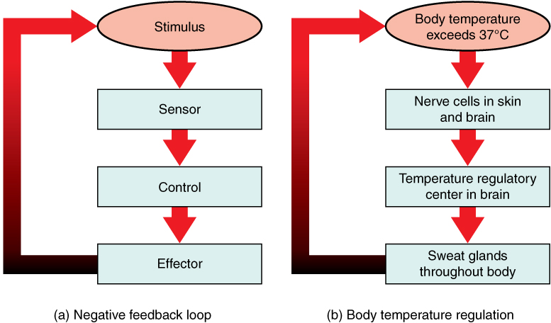
A negative feedback system requires at least four interacting components (Figure 1.3.2a):
- The stimulus is a change in the variable being regulated.
- The sensor is a receptor, often located in key places throughout the body and capable of detecting the stimulus and sending information to the control center.
- The control center receives information from the sensor and compares the incoming data to the normal range. If the stimulus is outside the normal range, then the control center directs the activity of an effector.
- The effector is an organ, gland, or muscle that responds to the signal from the control center in order to modify the variable.
.
In order to set the system in motion, a stimulus must drive a physiological parameter beyond its normal range (that is, beyond homeostasis). This stimulus is “heard” by a specific sensor. For example, in the control of blood glucose, specific endocrine cells in the pancreas detect excess glucose (the stimulus) in the bloodstream. These pancreatic beta cells respond to the increased level of blood glucose by releasing the hormone (insulin) into the bloodstream. The insulin signals skeletal muscle fibers, fat cells (adipocytes), and liver cells to take up the excess glucose, removing it from the bloodstream. As glucose concentration in the bloodstream drops, the decrease in concentration—the actual negative feedback—is detected by pancreatic alpha cells, and insulin release stops. This prevents blood sugar levels from continuing to drop below the normal range.
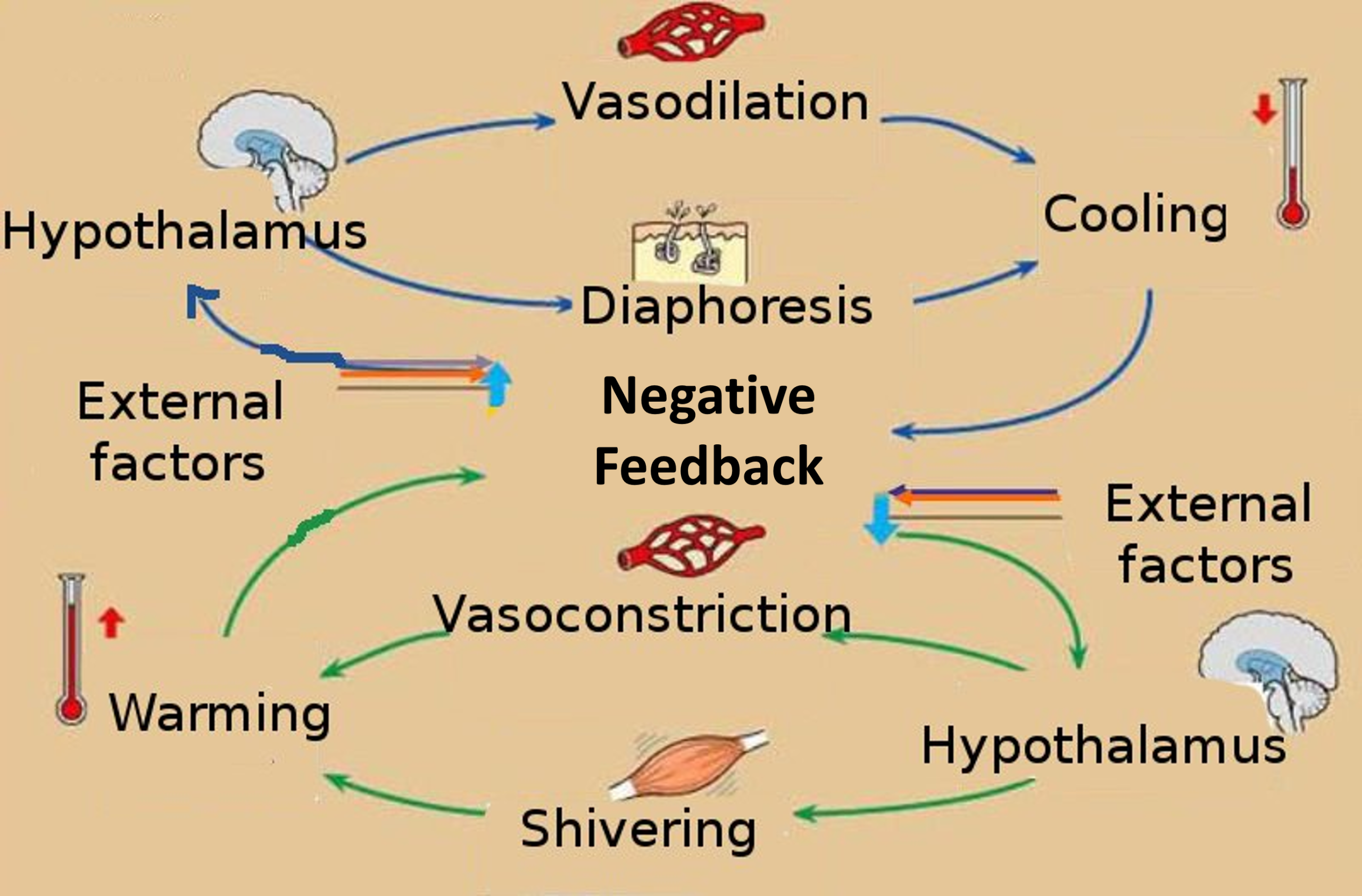
Humans have a similar temperature regulation feedback system that works by promoting either heat loss or heat gain (Figure 1.3.2b and Figure 1.3.3). When the brain’s temperature regulation center receives data from the sensors indicating that the body’s temperature exceeds its normal range, it stimulates a cluster of brain cells referred to as the “heat-loss center.” This stimulation has three major effects:
- Blood vessels in the skin begin to dilate, allowing more blood from the body core to flow to the surface of the skin and allowing the heat to radiate into the environment.
- As blood flow to the skin increases, sweat glands are activated to increase their output. As the sweat evaporates from the skin surface into the surrounding air, it takes heat with it.
- The depth of respiration increases, and a person may breathe through an open mouth instead of through the nasal passageways. This further increases heat loss from the lungs.
In contrast, activation of the brain’s heat-gain center by exposure to cold reduces blood flow to the skin, and blood returning from the limbs is diverted into a network of deep veins. This arrangement traps heat closer to the body core and restricts heat loss. If heat loss is severe, the brain triggers an increase in random signals to skeletal muscles, causing them to contract, producing shivering. The muscle contractions of shivering release heat while using up adenosine triphosphate (ATP). The brain triggers the thyroid gland in the endocrine system to release thyroid hormone, which increases metabolic activity and heat production in cells throughout the body. The brain also signals the adrenal glands to release epinephrine (adrenaline), a hormone that causes the breakdown of glycogen into glucose, which can be used as an energy source. The breakdown of glycogen into glucose also results in increased metabolism and heat production.

Interactive: Follow the QR code above to learn how the kidneys function in maintaining water balance.
Positive Feedback
Positive feedback intensifies a change in the body’s physiological condition rather than reversing it. A deviation from the normal range results in more change, and the system moves farther away from the normal range. Positive feedback in the body is normal only when there is a definite end point. Childbirth and the body’s response to blood loss are two examples of positive feedback loops that are normal but are activated only when needed.
Childbirth at full term is an example of a situation in which the maintenance of the existing body state is not desired. Enormous changes in the mother’s body are required to expel the baby at the end of pregnancy. The events of childbirth, once begun, must progress rapidly to a conclusion or the life of the mother and the baby are at risk. The extreme muscular work of labor and delivery are the result of a positive feedback system (Figure 1.3.4).
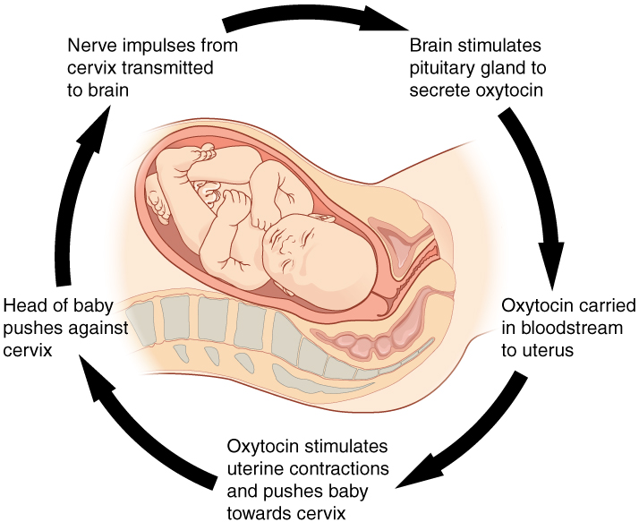
The first contractions of labor (the stimulus) push the baby toward the cervix (the lowest part of the uterus). The cervix contains stretch-sensitive nerve cells that monitor the degree of stretching (the sensors). These nerve cells send messages to the brain, which in turn causes the pituitary gland at the base of the brain to release the hormone oxytocin into the bloodstream. Oxytocin causes stronger contractions of the smooth muscles in of the uterus (the effectors), pushing the baby further down the birth canal. This causes even greater stretching of the cervix. The cycle of stretching, oxytocin release, and increasingly more forceful contractions stops only when the baby is born. At this point, the stretching of the cervix halts, stopping the release of oxytocin.A second example of positive feedback centers on reversing extreme damage to the body. Following a penetrating wound, the most immediate threat is excessive blood loss. Less blood circulating means reduced blood pressure and reduced perfusion (penetration of blood) to the brain and other vital organs. If perfusion is severely reduced, vital organs will shut down and the person will die. The body responds to this potential catastrophe by releasing substances in the injured blood vessel wall that begin the process of blood clotting. As each step of clotting occurs, it stimulates the release of more clotting substances. This accelerates the processes of clotting and sealing off the damaged area. Clotting is contained in a local area based on the tightly controlled availability of clotting proteins. This is an adaptive, life-saving cascade of events.
Section Review
Homeostasis is the activity of cells throughout the body to maintain the physiological state within a narrow range that is compatible with life. Homeostasis is regulated by negative feedback loops and, much less frequently, by positive feedback loops. Both have the same components of a stimulus, sensor, control center, and effector; however, negative feedback loops work to prevent an excessive response to the stimulus, whereas positive feedback loops intensify the response until an end point is reached.
Review Questions
Critical Thinking Questions
Glossary
- control center
- part of a feedback system that receives information from sensors and directs effectors to respond to stimuli
- dynamic equilibrium
- a state in which small changes in physiological variables are constantly balanced to maintain homeostasis
- effector
- an organ, gland, or muscle that acts in response to signals from a control center to restore homeostasis
- homeostasis
- the maintenance of a stable internal environment in the body
- negative feedback
- a mechanism that reverses a deviation from the set point to maintain homeostasis
- parameter
- a measurable physiological variable, such as temperature or blood pressure, regulated by feedback systems
- positive feedback
- a mechanism that intensifies a change in a physiological condition and moves the system farther from the normal range
- receptor
- a sensor that detects changes in physiological variables and sends information to the control center
- sensor
- a receptor that monitors internal conditions and responds to stimuli such as stretching or temperature changes
- set point
- the target physiological value around which a normal range fluctuates
- stimulus
- a change in the internal environment that activates a feedback loop
Glossary Flashcards
This work, Human Physiology, is adapted from Anatomy & Physiology by OpenStax, licensed under CC BY. This edition, with revised content and artwork, is licensed under CC BY-SA except where otherwise noted.
Images from Anatomy & Physiology by OpenStax are licensed under CC BY except where otherwise noted.
Access the original for free at OpenStax.
Image Descriptions
Figure 1.3.1. This diagram illustrates a negative feedback control system for maintaining homeostasis in biological organisms. The system consists of several key components that work together in a continuous loop. A sensor detects and measures the current value of the regulated variable (X), which is then sent to an error detector that compares this measurement against a set point (Y) to calculate the error signal (X,Y). This error signal is processed by a controller, which generates appropriate control signals that are sent to effectors. The effectors execute these signals to produce a physiological response that adjusts the regulated variable, which then feeds back to the sensor to complete the loop. The diagram is divided by a horizontal dotted line separating the external environment (above) from the internal environment (below). Two types of disturbances can affect this system: external disturbances, which are changes in external conditions that impact the internal environment, and internal disturbances, which are changes in the organism’s structure or function that affect the regulated variable’s magnitude. This represents the fundamental mechanism by which biological systems maintain stable internal conditions despite external and internal perturbations, a process known as homeostatic regulation. [Return to Figure 1.3.1]
Figure 1.3.2. This diagram compares a general negative feedback loop system with a specific example of body temperature regulation in biological organisms. Panel (a) shows the abstract negative feedback loop model, where a stimulus triggers a sensor that sends information to a control center, which then activates an effector to produce a response. A feedback arrow loops back from the effector to the stimulus, completing the cycle and indicating that the effector’s response works to counteract or reduce the initial stimulus. Panel (b) illustrates this same principle applied to body temperature regulation, where the stimulus is body temperature exceeding 37°C. This elevated temperature is detected by nerve cells in the skin and brain, which serve as the sensors. These nerve cells send signals to the temperature regulatory center in the brain, which acts as the control center. The brain then activates sweat glands throughout the body as the effectors, which produce sweat to cool the body down. The feedback arrow shows that this cooling response works to reduce the elevated body temperature back toward the normal set point, demonstrating how negative feedback maintains homeostasis by counteracting deviations from the optimal physiological state. [Return to Figure 1.3.2]
Figure 1.3.3. This diagram illustrates two complementary negative feedback loops that regulate body temperature in response to both heat and cold. The upper loop (shown in blue) demonstrates the body’s response to overheating, where the hypothalamus detects elevated temperature and triggers two cooling mechanisms: vasodilation, which widens blood vessels to increase heat loss through the skin, and diaphoresis, which is the production of sweat that cools the body through evaporation. As the body cools down, this information feeds back to the hypothalamus, which reduces these cooling responses. The lower loop (shown in green) shows the body’s response to cold, where the hypothalamus detects decreased temperature and initiates two warming mechanisms: vasoconstriction, which narrows blood vessels to reduce heat loss from the skin, and shivering, which generates heat through involuntary muscle contractions. As the body warms up, this feeds back to the hypothalamus, which then decreases these warming responses. Both loops are influenced by external factors such as environmental temperature, which can trigger either pathway. The diagram emphasizes that these are negative feedback systems because each response counteracts the initial stimulus, working to return body temperature to its optimal set point and maintain homeostasis. [Return to Figure 1.3.3]
Figure 1.3.4. This diagram shows the steps of a positive feedback loop as a series of stepwise arrows looping around a diagram of an infant within the uterus of a pregnant woman. Initially the head of the baby pushes against the cervix, transmitting nerve impulses from the cervix to the brain. Next the brain stimulates the pituitary gland to secrete oxytocin which is carried in the bloodstream to the uterus. Finally, the oxytocin simulates uterine contractions and pushes the baby harder into the cervix. As the head of the baby pushes against the cervix with greater and greater force, the uterine contractions grow stronger and more frequent. This mechanism is a positive feedback loop. [Return to Figure 1.3.4]
Report an Error
Did you find an error, typo, broken link, or other problem in the text? Please follow this link to the error reporting form to submit an error report to the authors.
