Chapter 11. The Cardiovascular System: Blood
11.0 Introduction
Chapter Objectives
After studying this chapter, you will be able to:
- Identify the primary functions of blood, its fluid and cellular components, and its characteristics
- Describe the formation of the formed element components of blood
- Compare and contrast the morphological features, relative size, abundance, and major functions of the three main formed elements of blood: erythrocytes (red blood cells), leukocytes (white blood cells), and thrombocytes (platelets)
- Compare and contrast how and where erythrocytes and leukocytes are produced
- Describe the process of hemostasis
- Explain the significance of AB and Rh blood groups in blood transfusions
- Discuss a variety of blood disorders
Single-celled organisms do not need blood. They obtain nutrients directly from and excrete wastes directly into their environment. The human organism cannot do that. Our large, complex bodies need blood to deliver nutrients to and remove wastes from our trillions of cells. The heart pumps blood throughout the body in a network of blood vessels. Together, these three components—blood, heart, and vessels—make up the cardiovascular system. This chapter focuses on the medium of transport: blood.
This work, Human Physiology, is adapted from Anatomy & Physiology by OpenStax, licensed under CC BY. This edition, with revised content and artwork, is licensed under CC BY-SA except where otherwise noted.
Images from Anatomy & Physiology by OpenStax are licensed under CC BY except where otherwise noted.
Access the original for free at OpenStax.
Image Descriptions
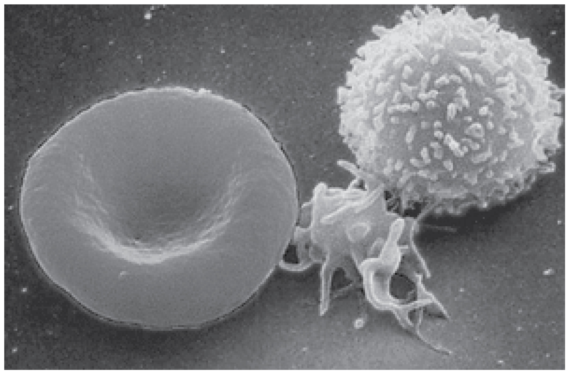
Figure 11.0.This is a scanning electron microscope (SEM) image displaying three types of human blood cells against a dark background. On the left is a red blood cell (erythrocyte), appearing as a smooth, round, biconcave disc with a characteristic depressed center. On the right is a white blood cell (leukocyte), distinguished by its spherical shape and heavily textured, bumpy surface covered in numerous protrusions. Between these two larger cells are several small, irregularly shaped platelets (thrombocytes) that appear as flat fragments with extending projections. The grayscale image shows the distinct morphological differences between these blood components in high magnification detail. [Return to Figure 11.0]
Report an Error
Did you find an error, typo, broken link, or other problem in the text? Please follow this link to the error reporting form to submit an error report to the authors.
