Chapter 13. The Urinary System
13.1 Internal and External Anatomy of the Kidney and Renal Blood Supply
Learning Objectives
By the end of this section, you will be able to:
- diagram or describe the structures and regions in a cross-sectional diagram of a kidney; and
- list the main blood vessels associated with the kidneys and nephrons and describe the direction of blood flow through these vessels.
Organs of the Urinary System
Urine produced by each kidney exits the kidney via a ureter, and the ureters deliver urine into the urinary bladder. The urinary bladder stores urine. Upon micturition (urination), urine exits the body via the urethra. In males, the urethra by which semen exits the body during ejaculation.
External Anatomy of the Kidney
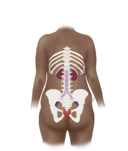
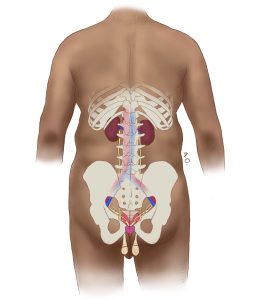
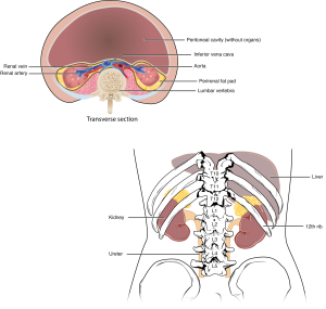
The paired kidneys lie on either side of the spine in the retroperitoneal space between the parietal peritoneum and the posterior abdominal wall, well protected by muscle, fat, and ribs. The left kidney is located at about the T12 to L3 vertebrae, whereas the right is lower due to slight displacement by the liver. Upper portions of the kidneys are somewhat protected by the eleventh and twelfth ribs (Figure 13.1.2). Each kidney weighs about 125 to 175 g in males and 115 to 155 g in females. They are about 11 to 14 cm in length, 6 cm wide, and 4 cm thick, and are directly covered by a fibrous capsule composed of dense, irregular connective tissue that helps to hold their shape and protect them. This capsule is covered by a shock-absorbing layer of adipose tissue called the renal fat pad, which in turn is encompassed by a tough renal fascia. The fascia and, to a lesser extent, the overlying peritoneum serve to firmly anchor the kidneys to the posterior abdominal wall in a retroperitoneal position. The adrenal glands, explored further in Chapter 17, are located on the superior aspects of each kidney.
Internal Anatomy of the Kidney
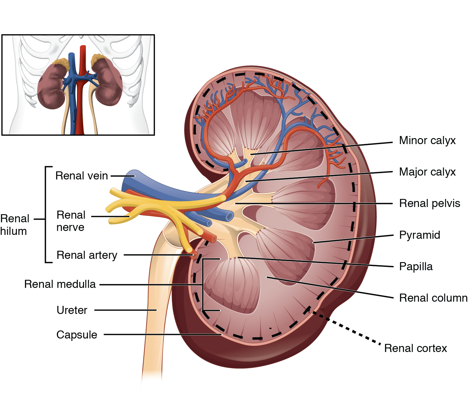
A frontal section through the kidney reveals an outer region called the renal cortex and an inner region called the renal medulla (Figure 13.1.3). The renal columns are extensions of the cortex that radiate downward through the medulla to separate the most characteristic features of the medulla, the renal pyramids. The renal columns also serve to divide the kidney into 6 to 8 lobes and provide a supportive framework for vessels that enter and exit the cortex.
Renal Pyramids, Major and Minor Calyces, and Renal Hilum
The renal pyramids are cone-shaped masses of tissue in the renal medulla. They are striated in appearance because they contain nephron collecting ducts as well as the nephron loops of juxtamedullary nephrons. The base of each pyramid faces the cortex, while the apex of each pyramid, the papilla, faces inward. The papillae drain urine from the collecting ducts to the calyces of the kidney for excretion.
Each renal papilla drains into a collecting pool called a minor calyx; several minor calyces connect to form a major calyx; all major calyces lead to the single renal pelvis, which is continuous with the ureter.
The renal hilum is the entry and exit site for structures servicing the kidneys: vessels, nerves, lymphatics, and ureters.
Blood Supply to the Kidneys
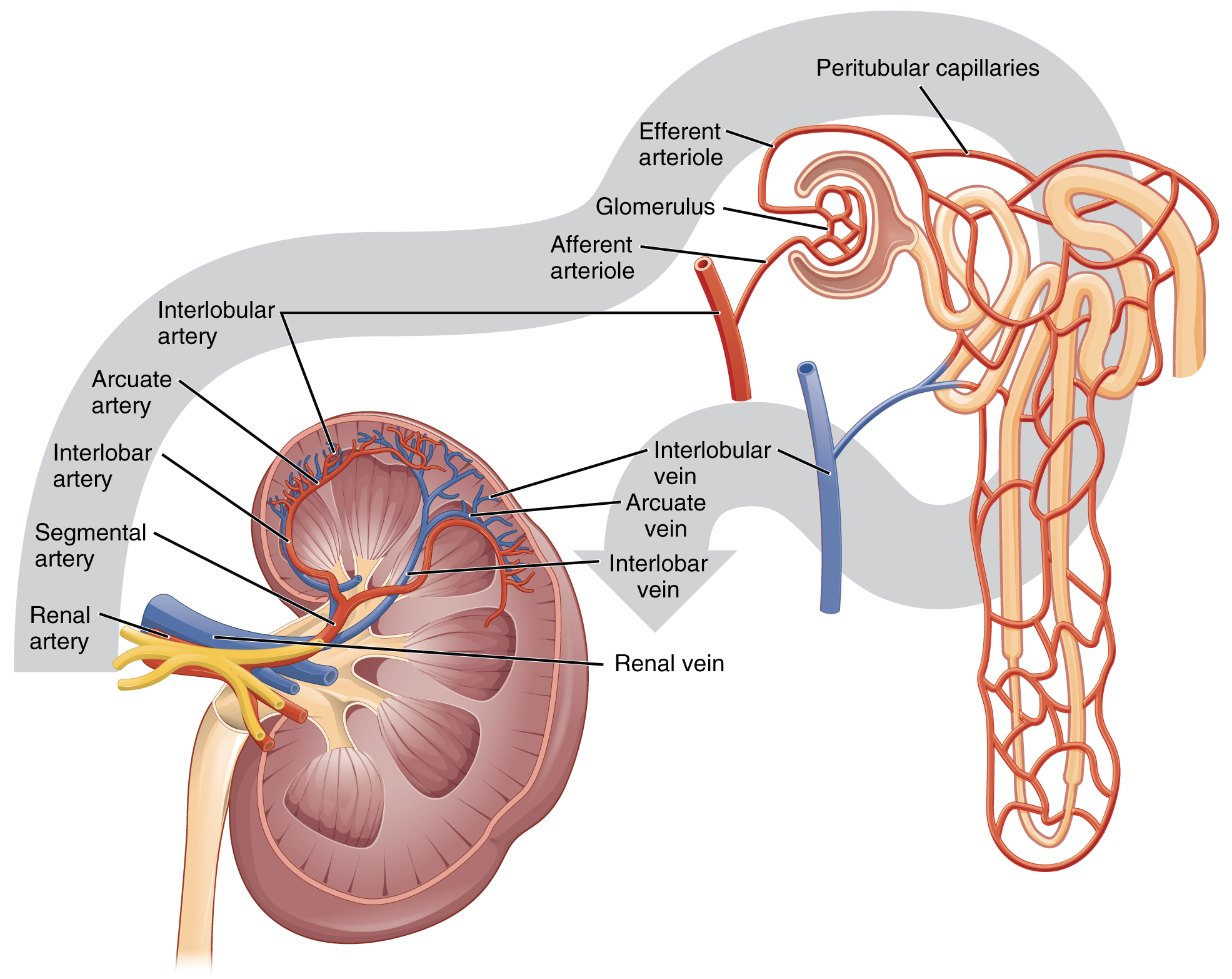
The kidneys are well vascularized and receive about 25% of the cardiac output at rest. Blood enters each kidney via a renal artery that branches directly from the descending aorta.
Once in the kidney, each renal artery divides into a five segmental arteries, which eventually further branch into interlobar arteries that pass through the renal columns to reach the cortex (Figure 13.1.4). The interlobar arteries, in turn, branch into arcuate arteries, cortical radiate arteries, and then into afferent arterioles. Each afferent arteriole delivers blood to a microscopic blood vessels blood are where blood enters the entry points the vessels that deliver blood to nephrons. Nephrons are the “functional units” of the kidneys; there are about 1.3 million nephrons in each kidney and they function to filter the blood. The detailed anatomy and functions of nephrons are described in more detail in subsequent chapters. Here we present an overview of the blood vessels important to delivering blood to each nephron.
Nephron Blood Flow
The afferent arterioles deliver blood into a modified capillary bed called the glomerulus which is a component of the “functional unit” of the kidney called the nephron. Once the nephrons have filtered the blood, renal veins return blood directly to the inferior vena cava. A portal system is formed when the blood flows from the glomerulus to the efferent arteriole through a second capillary bed, the peritubular capillaries (and vasa recta), surrounding the proximal and distal convoluted tubules and the loop of Henle. Most water and solutes are recovered by this second capillary bed. This filtrate is processed and finally gathered by collecting ducts that drain into the minor calyces, which merge to form major calyces; the filtrate then proceeds to the renal pelvis and finally the ureters.
External Website
For a review of the direction of blood flow to the kidneys and nephrons, watch the video below. Click here to access “Kidney Blood Supply – Blood Flow to, Through, and Away from Kidneys” by Interactive Biology (July 6, 2021) in a separate tab. Note that the nephron shown in the video is a cortical nephron. Types of nephrons will be discussed in the next section of this chapter.
Section Review
As noted previously, the structure of the kidney is divided into two principle regions—the outer cortex and the central medulla. The two kidneys receive about 25% of cardiac output. They are protected in the retroperitoneal space by the renal fat pad and overlying ribs and muscle. Ureters, blood vessels, lymph vessels, and nerves enter and leave at the renal hilum. The renal arteries arise directly from the aorta, and the renal veins drain directly into the inferior vena cava. Kidney function is derived from the actions of about 1.3 million nephrons per kidney; these are the “functional units” of the kidney. A capillary bed, the glomerulus, filters the blood and the filtrate is captured by the Bowman’s capsule. A portal system is formed when the blood flows through a second capillary bed surrounding the proximal and distal convoluted tubules and the loop of Henle. Most water and solutes are recovered by this second capillary bed. This filtrate is processed and finally gathered by collecting ducts that drain into the minor calyces, which merge to form the major calyces; the filtrate then proceeds to the renal pelvis and finally the ureters.
Review Questions
Critical Thinking Questions
Glossary
- afferent arteriole
- arteriole carrying blood into the glomerulus
- efferent arteriole
- arteriole carrying blood from the glomerulus to the capillary beds around the convoluted tubules and loop of Henle
- glomerulus
- capillary bed surrounded by Bowman’s capsule; filters the blood based on size
- nephrons
- functional units of the kidney that carry out all filtration and modification to produce urine; consist of renal corpuscles
- renal medulla
- inner region of kidney containing the renal pyramids
- peritubular capillaries
- capillary bed that surrounds the proximal and distal convoluted tubules and the loop of Henle in cortical nephrons
- renal cortex
- outer part of kidney
- renal papillae
- medullary area of the renal pyramids where collecting ducts empty urine into the minor calyces
- renal pyramids
- cone-shaped area in the medulla of the kidney containing collecting ducts and the loops of Henle of juxtamedullary nephrons
- ureters
- tubular structures that transport urine from kidneys to the urinary bladder
- urethra
- tubular structure by which urine leaves the urinary bladder and is excreted from the body during urination
- urinary bladder
- organ that stores urine
Glossary Flashcards
This work, Human Physiology, is adapted from Anatomy & Physiology by OpenStax, licensed under CC BY. This edition, with revised content and artwork, is licensed under CC BY-SA except where otherwise noted.
Images from Anatomy & Physiology by OpenStax are licensed under CC BY except where otherwise noted.
Access the original for free at OpenStax.
Report an Error
Did you find an error, typo, broken link, or other problem in the text? Please follow this link to the error reporting form to submit an error report to the authors.
