Chapter 13. The Urinary System
13.2 Microscopic Anatomy of the Kidney: Anatomy of the Nephron
Learning Objectives
By the end of this section, you will be able to:
- list the main parts of the nephron tubule and describe the order in which filtrate passes through them;
- compare and contrast cortical nephrons and juxtamedullary nephrons;
- describe the components of and function of the renal corpuscle;
- describe the structure of the filtration membrane;
- describe the structure and function of the juxtaglomerular apparatus; and
- describe the histology of and functional significance of the proximal convoluted tubule, loop of Henle, distal convoluted tubule, and collecting ducts.
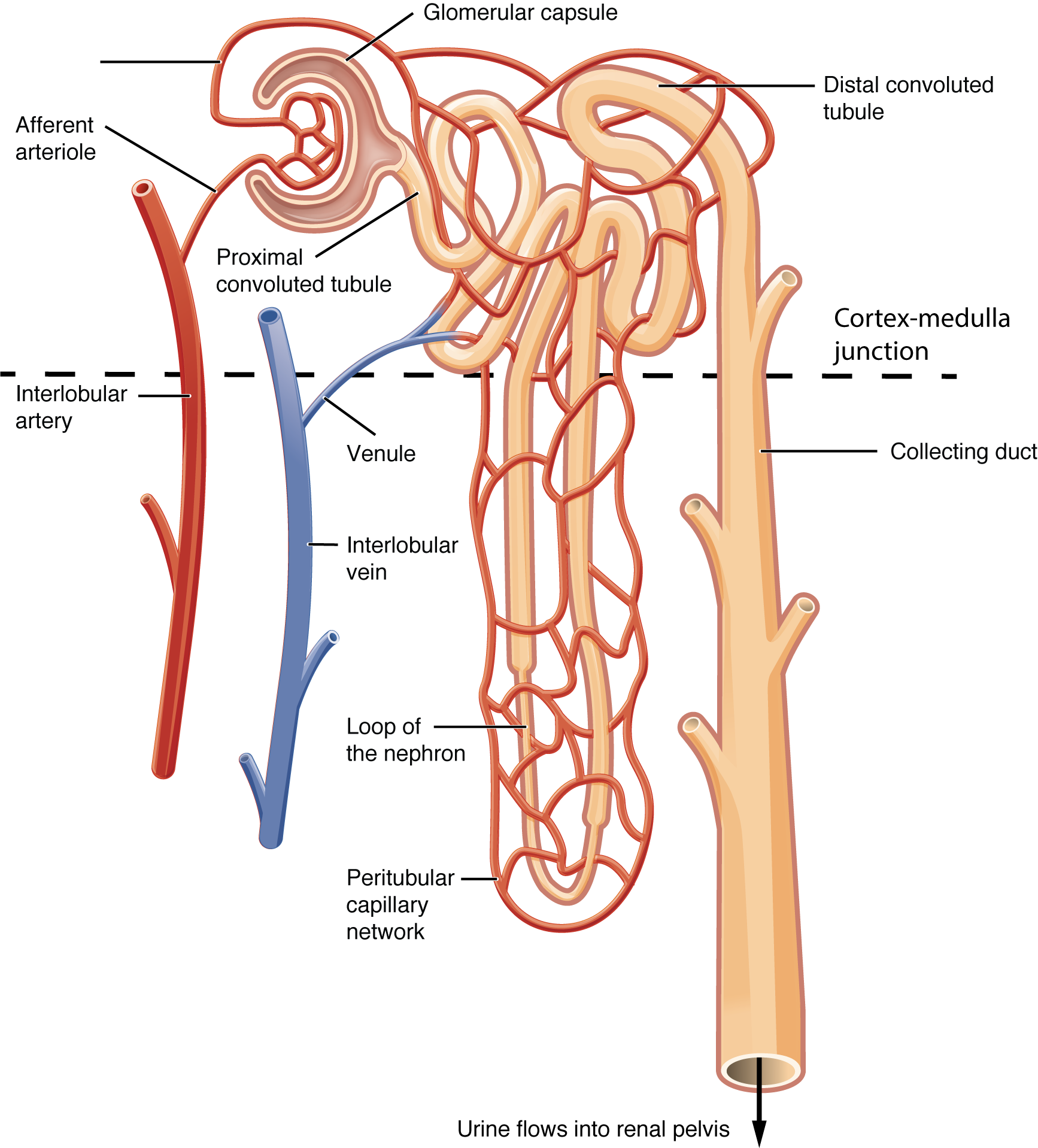
As mentioned in the previous section, the nephron is the functional unit of the kidney. It is functionally compartmentalized: it is a long tubule with different parts that have distinct functions. Generally speaking, nephrons rid the blood of toxins and balance the constituents of the plasma to homeostatic set points.
- Glomerular capsule (also called Bowman’s capsule)
- Proximal convoluted tubule (PCT)
- Loop of Henle (also called the nephron loop)
- Distal convoluted tubule (DCT)
- Collecting duct

Microanatomy of the Nephron
Renal Corpuscle
As discussed earlier, the renal corpuscle consists the glomerulus and the glomerular capsule. The glomerulus is a high pressured, fenestrated capillary with large holes (fenestrations) between the endothelial cells. The glomerular capsule captures the filtrate created by the glomerulus and directs this filtrate to the PCT. The outermost part of glomerular capsule is a simple squamous epithelium. It transitions over the glomerulus as uniquely shaped cells (podocytes) with finger-like arms (pedicels) that cover the glomerular capillaries (Figure 13.2.3). A thin basement membrane lies between the glomerular endothelium and the podocytes. The pedicels interdigitate to form filtration slits, leaving small gaps that form a sieve. As blood passes through the glomerulus, 10% to 20% of the plasma filters out of the fenestrations, through the basement membrane and between these sieve-like fingers to be captured by the glomerular capsule and funneled to the PCT. These features comprise the filtration membrane.
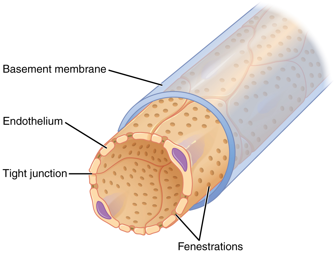
The filtration membrane prevents passage of blood cells, large proteins, and most negatively charged particles but allows most other constituents through. These substances cross readily if they are less than 4 nm in size and most pass freely up to 8 nm in size. Negatively charged particles have difficulty leaving the blood because the proteins associated with the filtration membrane are negatively charged, so they tend to repel negatively charged substances and allow positively charged substances to pass more readily. Asa result, the filtrate that does not contain cells or large proteins, and has a slight predominance of positively charged substances.
Proximal Convoluted Tubule (PCT)
Filtered fluid collected by the glomerular capsule enters into the PCT. Simple cuboidal cells form this tubule with prominent microvilli on the luminal surface, forming a brush border. These microvilli create a large surface area to maximize the reabsorption from and secretion of solutes into the filtrate. Reabsorption and secretion are the main functions of the PCT.
Loop of Henle
The descending and ascending limbs of the loop of Henle (sometimes referred to as the nephron loop) are continuations of the same tubule. They run adjacent and parallel to each other after making a hairpin turn at the deepest point of their descent. The descending limb of the loop of Henle consists of an initial short, thick portion and long, thin portion, whereas the ascending loop consists of an initial short, thin portion followed by a long, thick portion. The descending and ascending thick portions consist of simple cuboidal epithelium. The descending and ascending thin portions consists of simple squamous epithelium. These distinct histologies result in different permeabilities for solutes and water in the different portions of the loop. Reabsorption is the main function of the loop of Henle.
Distal Convoluted Tubule (DCT)
The DCT, like the PCT, is formed by simple cuboidal epithelium, but it is shorter than the PCT. These cells are not as active as those in the PCT and there are fewer microvilli on the apical surface. However, the DCT still carries out reabsorption and secretion of select substances according to the levels of certain hormones.
Collecting Ducts
The collecting ducts are continuous with the nephron but not technically part of it. In fact, each duct collects filtrate from several nephrons for final modification. Collecting ducts merge as they descend deeper in the medulla to form about 30 terminal ducts that empty at a papilla. They are lined with simple cuboidal epithelium. Cells called principal cells, found in the collecting duct and in the late portion of the DCT, enable water reabsorption in response to the hormone ADH. ADH is discussed later in this chapter.
Juxtaglomerular Apparatus (JGA)
A second function of the macula densa cells is to regulate renin release from the juxtaglomerular cells (granular cells) of the afferent arteriole.

Section Review
The functional unit of the kidney, the nephron, consists of the renal corpuscle, PCT, loop of Henle, and DCT. Cortical nephrons have short loops of Henle, whereas juxtamedullary nephrons have long loops of Henle extending into the medulla. About 15% of nephrons are juxtamedullary. The glomerulus is a capillary bed that filters blood principally based on particle size. A filtration membrane is formed by the fused basement membranes of the podocytes and the glomerular capillary endothelial cells. The formed filtrate enters the glomerular capsule followed by the PCT.
Specialized cells in the juxtaglomerular apparatus (JGA) produce paracrine signals to regulate blood flow and filtration rates of the glomerulus. Other JGA cells produce the enzyme renin, which plays a central role in blood pressure regulation. The filtrate enters the PCT where reabsorption and secretion of several substances occur. The descending and ascending limbs of the loop of Henle consist of thick and thin segments. Reabsorption and secretion continue in the DCT but to a lesser extent than in the PCT. Each collecting duct collects urine from several nephrons and functions to fine tune water reabsorption.
Review Questions
Critical Thinking Questions
Glossary
- brush border
- formed by microvilli on the surface of certain cuboidal cells; in the kidney it is found in the PCT; increases surface area for absorption in the kidney
- fenestrations
- small pores in a cell, allowing rapid filtration based on size
- filtration slits
- spaces formed by pedicels of podocytes; along with fenestrations, allow filtration to occur
- filtrate
- a plasma-like liquid (lacking cells and most proteins) formed during filtration
- juxtaglomerular apparatus (JGA)
- located at the juncture of the DCT and the afferent and efferent arterioles of the glomerulus; plays a role in the regulation of renal blood flow and GFR
- juxtaglomerular cells
- modified smooth muscle cells of the afferent arteriole; secrete renin in response to a drop in blood pressure; also called granular cells
- macula densa
- cells found in the part of the DCT forming the JGA; sense Na+ concentration in the forming urine
- pedicels
- finger-like projections of podocytes surrounding glomerular capillaries; interdigitate to form a filtration membrane
- podocytes
- cells forming finger-like processes; form the visceral layer of Bowman’s capsule; pedicels of the podocytes interdigitate to form a filtration membrane
- principal cell
- cell type found in the collecting duct and in the late portion of the DCT; under the control of ADH, are responsible for water reabsorption
Glossary Flashcards
This work, Human Physiology, is adapted from Anatomy & Physiology by OpenStax, licensed under CC BY. This edition, with revised content and artwork, is licensed under CC BY-SA except where otherwise noted.
Images from Anatomy & Physiology by OpenStax are licensed under CC BY except where otherwise noted.
Access the original for free at OpenStax.
Report an Error
Did you find an error, typo, broken link, or other problem in the text? Please follow this link to the error reporting form to submit an error report to the authors.
