Chapter 13. The Urinary System
13.7 Urine Composition, Transport, and Elimination
Learning Objectives
By the end of this section, you will be able to:
- describe the characteristics of a normal urine sample;
- explain how the integration of renal filtration, reabsorption, and secretion determines the volume of and composition of urine;
- identify the ureters, urinary bladder, and urethra and describe their function;
- define micturition and incontinence;
- describe voluntary and involuntary neural control of micturition; and
- explain the main difference in the roles of the internal and external urethral sphincters in micturition
Composition of Urine
As previously mentioned, the kidneys filter the entire plasma volume about 60 times each day. Once filtrate is formed, substances that need to be reclaimed back into the blood are reabsorbed, and substances that need to be eventually excreted in the urine stay in the filtrate and/or are added to the filtrate by secretion. Urine composition is variable (Table 13.4), depending on water intake and losses, nutrient intake, and other factors; however, blood cells and proteins are not normally found in the urine. Urine characteristics such as color and odor are rough descriptors of a person’s state of hydration. For example, if you exercise or work outside and sweat a great deal, your urine will be darker in color due to an increased urine concentration. Alternatively, a well hydrated person will have light or clear colored urine due to a more dilute urine (Figure 13.7.1).
| Characteristic | Normal Values |
|---|---|
| Color | Pale yellow to deep amber |
| Odor | Odorless |
| Volume | 800 to 2,000 mL/24 hour |
| pH | 4.5 to 8.0 |
| Specific gravity | 1.003 to 1.032 |
| Osmolarity | 40 to 1,350 mOsM |
| Urobilinogen | 0.1 to 1.8 mg/100 mL |
| White blood cells | 0 to 2 HPF (per high-power field of microscope) |
| Leukocyte esterase | None |
| Protein | None or trace |
| Bilirubin | <0.3 mg/100 mL |
| Ketones | None |
| Nitrites | None |
| Blood | None |
| Glucose | None |

The pH of the urine can vary more than 1,000-fold, from a normal low of 4.5 to a maximum of 8.0, depending on actions of specific cells of the kidney and on diet. Urine osmolarity ranges from a low of 50 to 100 mOsM to as high as 1,200 mOsM. The color of urine is determined mostly by the breakdown products of red blood cell destruction. The “heme” of hemoglobin is converted by the liver into water-soluble forms that can be excreted into the bile and indirectly into the urine. This yellow pigment is urochrome and its urine levels depend on several factors, namely hydration status. Urobilinogen is another byproduct of heme breakdown and normal levels range from 0.08 to 1.8 mg/dL.
Urine color may also be affected by certain foods like beets, berries, and fava beans. Dehydration produces darker, concentrated urine that may also possess the slight odor of ammonia. Ammonia (NH3) is a toxic byproduct of protein metabolism. It is formed as amino acids are deaminated by liver hepatocytes. That means that the amine group, NH2, is removed from amino acids as they are broken down. Most of the resulting ammonia is converted into urea by liver hepatocytes. Urea is not only less toxic but is also one of the main solutes contributing to the vertical osmotic gradient in the kidney interstitial fluid.
Urine volume varies considerably. The normal range is 800 to 2,000 mL per day. The kidneys must produce a minimum urine volume of about 400 mL per day to rid the body of wastes. Output below this level may be caused by severe dehydration or renal disease.
Nitrogenous Wastes in Urine
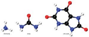
Nitrogenous wastes are produced by the breakdown of proteins during normal metabolism. Proteins are broken down into amino acids, which in turn are deaminated by having their nitrogen groups removed. Deamination converts the amino (NH2) groups into ammonia (NH3), ammonium ion (NH4+), urea, or uric acid (Figure 13.7.2). Ammonia is extremely toxic, so most of it is very rapidly converted into urea in the liver. Human urinary wastes typically contain primarily urea with small amounts of ammonium and very little uric acid.
Disorders of the Urinary Tract – Renal Calculi (Kidney Stones)
Urine Transport, Storage, and Elimination
All structures involved in the transport and storage of the urine are large enough to be visible to the naked eye. This transport and storage system not only stores the waste, but it protects the tissues from damage due to the wide range of pH and osmolarity of the urine.
Ureters
As urine is formed, it drains into the calyces of the kidney, which merge to form the funnel-shaped renal pelvis in the hilum of each kidney. The renal pelvis narrows to become the ureter of each kidney. As urine passes through the ureter, it does not passively drain into the bladder but rather is propelled by waves of peristalsis. As the ureters enter the pelvis, they pass laterally, hugging the pelvic walls. As they approach the bladder, they turn medially and join with the bladder wall obliquely (Figure 13.7.4.) This is important because it creates a physiological sphincter that allows urine into the bladder but prevents reflux of urine from the bladder back into the ureter. Children born lacking this oblique course of the ureter through the bladder wall are susceptible to “vesicoureteral reflux,” which dramatically increases their risk of serious UTI (urinary tract infection.) Pregnancy also increases the likelihood of reflux and UTI.
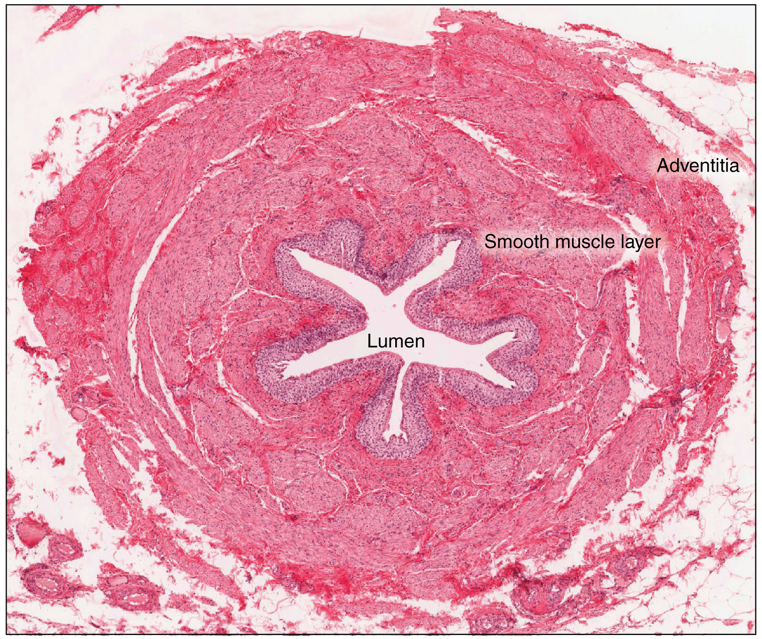
The ureters are approximately 30 cm long. The inner mucosa is lined with transitional epithelium (Figure 13.7.3) and scattered goblet cells that secrete protective mucus. The muscular layer of the ureter consists of longitudinal and circular smooth muscles that create the peristaltic contractions to move the urine into the bladder without the aid of gravity. Finally, a loose adventitial layer composed of collagen and fat anchors the ureters between the parietal peritoneum and the posterior abdominal wall.
Bladder
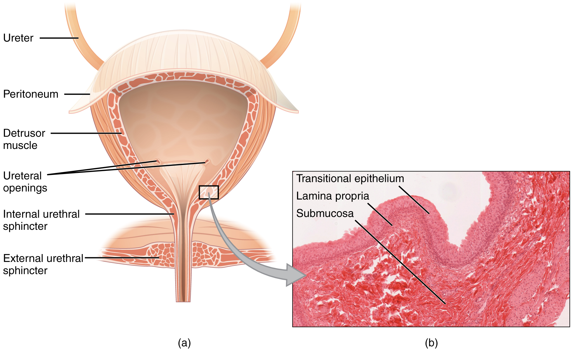
The urinary bladder collects urine from both ureters (Figure 13.7.4). The bladder lies posterior to the pubic bone and anterior to the rectum. The bladder is partially retroperitoneal (outside the peritoneal cavity) with its peritoneal-covered “dome” projecting into the abdomen when the bladder is distended with urine. When empty, the region of the bladder that does not collapse is called the trigone (Greek “tri-” meaning “triangle”), which is delineated by the opening of the ureters and the urethra, forming a triangular area.
External Website
View the University of Michigan WebScope here to explore the bladder tissue sample in greater detail.

The bladder is a highly distensible organ comprised of irregular crisscrossing bands of smooth muscle collectively called the detrusor muscle. The interior surface is made of transitional cellular epithelium that is structurally suited for the large volume fluctuations of the bladder. When empty, it resembles columnar epithelia, but when stretched, it “transitions” (hence the name) to a squamous appearance Figure 13.7.4). Normal bladder volumes in adults can range from about 50 mL when empty to 500 to 600 mL when full.
The detrusor muscle contracts with significant force in the young. The bladder’s strength diminishes with age, but voluntary contractions of abdominal skeletal muscles can increase intra-abdominal pressure to promote more forceful bladder emptying. Such voluntary contraction is also used in forceful defecation and childbirth.
Urethra
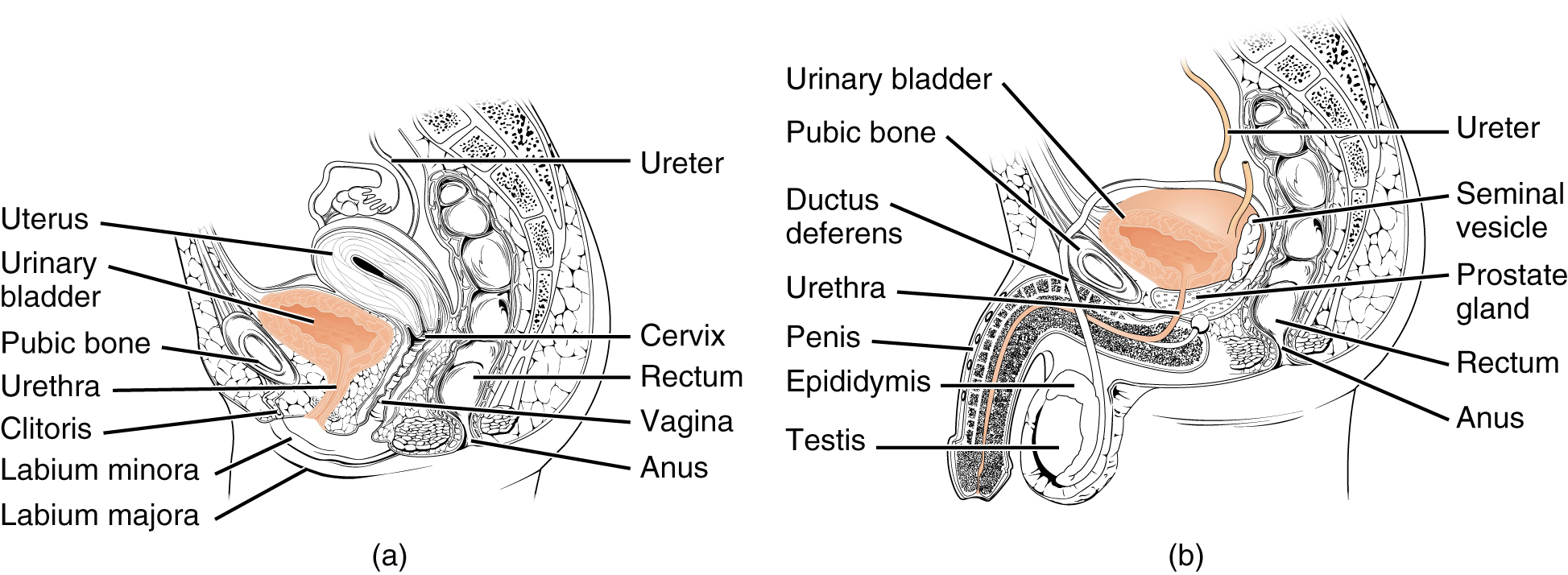
The urethra transports urine from the bladder to the outside of the body for disposal. The urethra is the only urologic organ that shows any significant anatomic difference between males and females. The urethra in females is about 3 to 4 cm in length, while in males it is approximately 20 cm long. All other urine transport structures are identical between females and males (Figure 13.7.5).
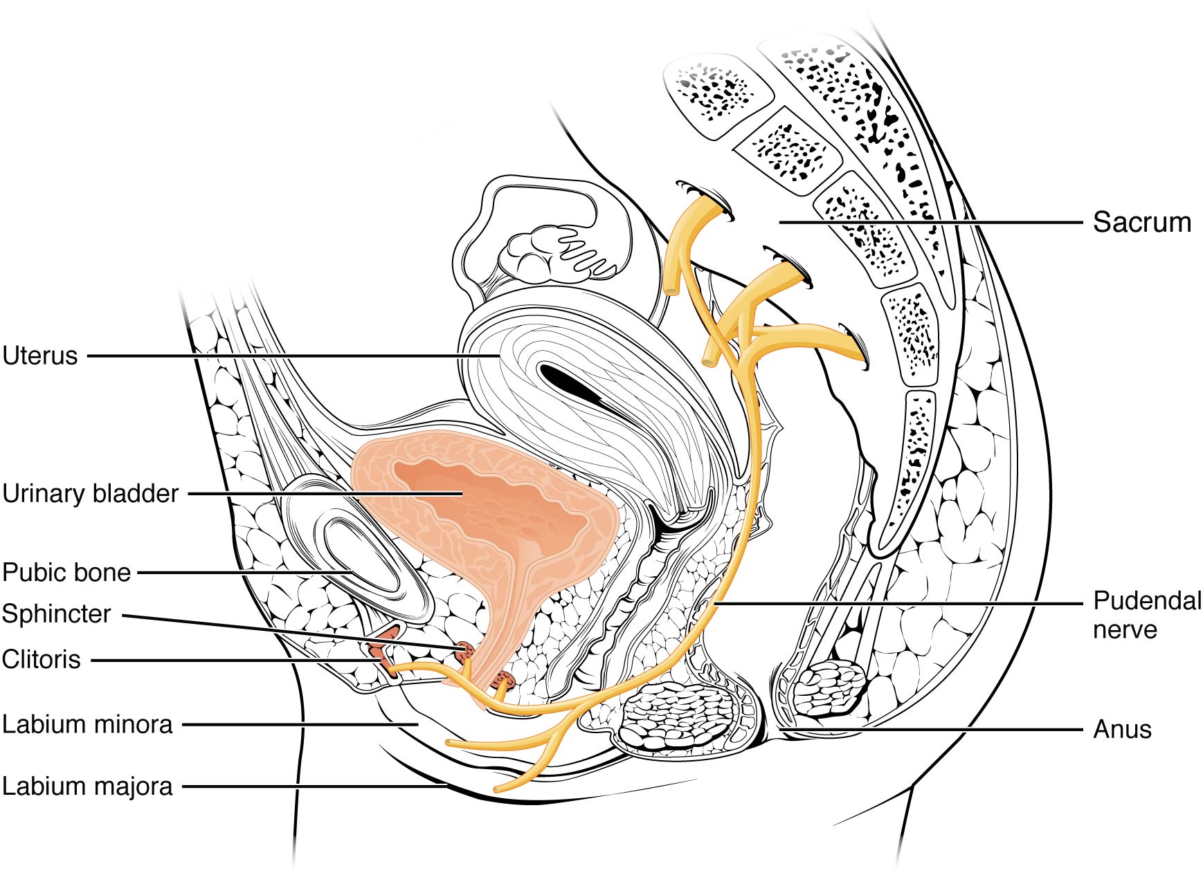
The urethra in both males and females begins inferior and central to the trigone. The urethra tracks posterior and inferior to the pubic symphysis (Figure 13.7.5). In both males and females, the proximal urethra is lined by transitional epithelium, whereas the terminal portion is a nonkeratinized, stratified squamous epithelium. In the male, pseudostratified columnar epithelium lines the urethra between these two cell types. Voiding is regulated by an involuntary autonomic nervous system-controlled internal urethral sphincter consisting of smooth muscle, and by the external urethral sphincter, which is comprised of skeletal muscle. Voluntary control of the external urethral sphincter is a function of the pudendal nerve. It arises in the sacral region of the spinal cord, traveling via the S2 to S4 nerves of the sacral plexus.
Disorders of the Urinary Tract – Urinary Tract Infection (UTI)
Urinary tract infections (UTIs) are bacterial infections of the kidneys, bladder, ureters, or urethra. They are usually caused by E. coli and other bacteria from the bowel or the bladder that enter the tract and proliferate. While UTIs do occur in males, they are more common in females for two reasons: 1) the shorter urethra in females means a shorter distance of travel for bacteria to reach the bladder; and 2) the female urethral opening is closer to the anus than in males, making it easier for fecal bacteria to enter the urinary tract. In addition, sexual activity can increase the risk of UTI in females since bacteria that collect in the vagina during intercourse can gain entry into the urinary tract through the nearby urethral opening. Nearly 50% of females will experience a UTI in their lifetime, a greater likelihood than in males.
Additionally, catheterization and any other condition that obstructs the flow of urine can lead to a UTI. For example, males with a swollen prostate have an increased likelihood of UTI: the enlarged prostate constricts the urethra and this can result in insufficient voiding of the bladder. The urine remaining in the bladder can accumulate bacteria and this growth can lead to a UTI.
Micturition Reflex
Micturition is the physiological term for urination or voiding. It results from an interplay of involuntary and voluntary actions by the internal and external urethral sphincters. When bladder volume reaches about 150 mL, an urge to void is sensed but is easily overridden. Voluntary control of urination relies on consciously preventing relaxation of the external urethral sphincter to maintain urinary continence. As the bladder fills, subsequent urges become harder to ignore.
Micturition is a result of stretch receptors in the bladder wall that transmit nerve impulses to the sacral region of the spinal cord to generate a spinal reflex. The resulting parasympathetic neural outflow causes contraction of the detrusor muscle and relaxation of the involuntary internal urethral sphincter. At the same time, the spinal cord inhibits somatic motor neurons, resulting in the relaxation of the skeletal muscle of the external urethral sphincter. The micturition reflex is active in infants but with maturity, children learn to override the reflex by asserting external sphincter control, thereby delaying voiding (potty training).
Nerves involved in the control of urination include the hypogastric, pelvic, and pudendal nerves. Voluntary micturition requires an intact spinal cord and functional pudendal nerve arising from the sacral micturition center (Figure 13.7.6). Since the external urethral sphincter is voluntary skeletal muscle, actions by somatic motor neurons maintain contraction (and thereby continence) during filling of the bladder. At the same time, sympathetic nervous activity via the hypogastric nerves suppresses contraction of the detrusor muscle. With further bladder stretch, afferent signals traveling over sacral pelvic nerves activate parasympathetic neurons. This activates efferent neurons to release acetylcholine at the neuromuscular junctions, producing detrusor contraction and bladder emptying.
Incontinence refers to the inability to prevent discharge of urine from the bladder. Causes of incontinence range from disruption of neural pathways mediating voluntary micturition, to less serious and more frequent causes such as sudden increases in bladder pressure. Examples of the latter include coughing or sneezing combined with impaired sphincter function. Incontinence can range from occasional, mild leakage to more frequent and severe loss of control.
Section Review
Urine composition varies depending on water intake and losses, nutrient intake, and other factors; however, blood cells and proteins are not normally found in the urine. Urine color is predominantly due to urochrome, a yellow pigment resulting from heme breakdown.
Nitrogenous wastes produced in protein metabolism are excreted in urine, with urea being the primary nitrogenous waste. Ammonium and uric acid are excreted in smaller amounts.
The urethra is the only urinary structure that differs significantly between males and females. This is due to the dual role of the male urethra in transporting both urine and semen.
Micturition is the process of voiding the urine and involves both involuntary and voluntary actions. Voluntary control of micturition requires a mature and intact sacral micturition center. It also requires an intact spinal cord.
Review Questions
Critical Thinking Questions
Glossary
- detrusor muscle
- smooth muscle in the bladder wall; fibers run in all directions to reduce the size of the organ when emptying it of urine
- external urethral sphincter
- skeletal muscle forms this sphincter; must be relaxed consciously to void urine
- internal urethral sphincter
- smooth muscle sphincter at the juncture of the bladder and urethra; relaxes as the bladder fills to allow urine into the urethra
- incontinence
- loss of ability to control micturition
- micturition
- also called urination or voiding
- retroperitoneal
- outside the peritoneal cavity; in the case of the kidney and ureters, between the parietal peritoneum and the abdominal wall
- sacral micturition center
- group of neurons in the sacral region of the spinal cord that controls urination; acts reflexively unless its action is modified by higher brain centers to allow voluntary urination
- trigone
- area at the base of the bladder marked by the two ureters in the posterior–lateral aspect and the urethral orifice in the anterior aspect; does not collapse when the bladder is empty
- urethra
- transports urine from the bladder to the outside environment
Glossary Flashcards
References
Cleveland Clinic. (2025, February 10). Urethra: Location, anatomy, Function & Conditions. Cleveland Clinic.
Cleveland Clinic. (2025c, June 2). What are kidney stones? Cleveland Clinic.
This work, Human Physiology, is adapted from Anatomy & Physiology by OpenStax, licensed under CC BY. This edition, with revised content and artwork, is licensed under CC BY-SA except where otherwise noted.
Images from Anatomy & Physiology by OpenStax are licensed under CC BY except where otherwise noted.
Access the original for free at OpenStax.
Report an Error
Did you find an error, typo, broken link, or other problem in the text? Please follow this link to the error reporting form to submit an error report to the authors.
