Chapter 14. Fluid, Electrolyte, and Acid-Base Balance
14.2 Fluid Balance: Maintaining Electrolyte Balance and ECF Volume
Learning Objectives
By the end of this section, you will be able to:
- state examples of important roles of sodium, potassium, chloride, bicarbonate, calcium, and phosphate;
- identify the following: the main ECF cation, the main ECF anion, and the main ICF cation;
- explain how sodium is the main electrolyte that determines ECF volume and body water distribution;
- state the disorders associated with abnormally high and low levels of sodium, potassium, chloride, calcium, and phosphate;
- describe how sodium, potassium, chloride, calcium, and phosphate are regulated;
- explain the roles of RAAS and ANP in indirectly regulating ECF volumer by regulating body sodium content;
- explain the role of RAAS in the long term regulation of blood pressure; and
- list three ways in which plasma pH is maintained.
Body fluids contain a variety electrolytes that perform a number of important functions. Some electrolytes assist in the transmission of electrical impulses along cell membranes in neurons and muscles. Others help to stabilize protein structures in enzymes. Still others aid in releasing hormones from endocrine glands. All of the electrolytes in plasma contribute to the ECF osmolarity that controls the movement of water between cells (ICF) and their environment (ECF).
Electrolytes in living systems include sodium, potassium, chloride, bicarbonate, calcium, phosphate, magnesium, copper, zinc, iron, manganese, molybdenum, copper, and chromium.
In terms of body functioning, six of the most important electrolytes are: sodium, potassium, chloride, bicarbonate, calcium, and phosphate. These six ions aid in nerve excitability, endocrine secretion, membrane permeability, buffering body fluids, and controlling the movement of fluids between compartments.
Electrolytes enter the body via the digestive tract. More than 90% of the calcium and phosphate that enters the body is incorporated into bones and teeth, with bone serving as a mineral reserve for these ions. In the event that calcium and phosphate are needed for other functions, bone tissue can be broken down to supply the blood and other tissues with these minerals. Phosphate is a normal constituent of nucleic acids; hence, blood levels of phosphate will increase whenever nucleic acids are broken down.
Excretion of electrolytes occurs mainly via in the urine, with lesser amounts lost in sweat and in feces. Excessive sweating may cause a significant loss, especially of sodium and chloride. Severe vomiting or diarrhea will also cause electrolyte loss, especially bicarbonate in cases of diarrhea, and H+ in cases of severe vomiting. Adjustments in respiratory and renal functions allow the body to regulate the levels of these ions in the ECF, and this is discussed in the next section.
Table 14.3 lists the reference values for plasma and urine for the six electrolytes addressed in this section. In a clinical setting, analysis of urine electrolyte levels requires a collection of urine across a 24-hour period, because the output of these ions can vary considerably over the course of a day. Urine values reflect the rates of excretion of these ions. Bicarbonate is not normally excreted in urine; instead, it is conserved by the kidneys and is an important buffer in the ECF.
| Name | Chemical Symbol | Plasma | Urine |
|---|---|---|---|
| Sodium | Na+ | 135.0 – 145.0 mM
(135.0 – 145.0 mEq/L) |
40.0 – 220.0 mEq/24 hr |
| Potassium | K+ | 3.5 – 5.5 mM
(3.5 – 5.5 mEq/L) |
25.0 – 125.0 mEq/24 hr |
| Chloride | Cl– | 97.0 – 105.0 mM
(97.0 – 105.0 mEq/L) |
110.0 – 250.0 mEq/24 hr |
| Bicarbonate | HCO3– | 22.0 – 26.0 mEq/L
(22.0 – 26.0 mM) |
N/A |
| Calcium
|
Ca++ | 4.3 – 5.3 mEq/L | Up to 7.5 mmol/24 hr |
| Phosphate | Two main forms: H2PO4 – and HPO42- | 0.8 – 1.5 mM | 400 – 1300 mg/24 hr, varies with diet |
Sodium
Sodium is the major cation of the extracellular fluid. It is the electrolyte that contributes the most to the osmotic pressure of the ECF. As discussed in the previous section, sodium plays a main role in determining ECF volume and body water distribution. People eating a typical Western diet, which is very high in NaCl, routinely take in 130 to 160 mmol/day of sodium, but humans require only 1 to 2 mmol/day. Diets higher in sodium are associated with a greater risk of developing hypertension, a major cause of stroke and heart disease. Excretion of sodium is accomplished primarily by the kidneys. Sodium is freely filtered through the glomerular capillaries of the kidneys, and although much of the filtered sodium is reabsorbed in the PCT, some remains in the filtrate and urine, and is normally excreted.
Hyponatremia is a lower-than-normal plasma sodium concentration, usually associated with excess water accumulation in the body (hypotonic hydration). An absolute loss of sodium may be due to a decreased intake of the ion coupled with its continual excretion in the urine. An abnormal loss of sodium from the body can result from several conditions, including excessive sweating, vomiting, or diarrhea; the use of diuretics; excessive production of urine, which can occur in diabetes; and certain types of metabolic acidosis. A relative decrease in blood sodium can occur because of an imbalance of sodium in one of the body’s other fluid compartments, like IF, or from a dilution of sodium due to water retention related to edema or congestive heart failure. At the cellular level, hyponatremia results in increased entry of water into cells by osmosis, because the concentration of solutes within the cell exceeds the concentration of solutes in the now-diluted ECF. The excess water causes swelling of the cells; the swelling of red blood cells—decreasing their oxygen-carrying efficiency and making them potentially too large to fit through capillaries—along with the swelling of neurons in the brain can result in brain damage or even death.
Hypernatremia is an abnormal increase of blood sodium. It can result from water loss from the blood, resulting in the hemoconcentration of all blood constituents. This can lead to neuromuscular irritability, convulsions, CNS lethargy, and coma. Hormonal imbalances involving ADH and aldosterone may also result in higher-than-normal sodium values.
Potassium
Potassium is the major intracellular cation. It helps establish the resting membrane potential in neurons and muscle fibers after membrane depolarization and action potentials. In contrast to sodium, potassium has very little effect on osmotic pressure. The low level of potassium in blood is due to the sodium-potassium pumps in cell membranes, which maintain the normal potassium concentration gradient between the ICF and ECF. The recommendation for daily intake/consumption of potassium is 4,700 mg. Potassium is excreted, both actively and passively via the nephrons, especially the DCTs and collecting ducts.
Hypokalemia is an abnormally low potassium blood level. Similar to the situation with hyponatremia, hypokalemia can occur because of either an absolute reduction of potassium in the body or a relative reduction of potassium in the blood due to the redistribution of potassium. An absolute loss of potassium can arise from decreased intake, frequently related to starvation. It can also come about from vomiting, diarrhea, or alkalosis. Hypokalemia can cause metabolic acidosis, CNS confusion, and cardiac arrhythmias.
Some insulin-dependent diabetic patients experience a relative reduction of potassium in the blood from the redistribution of potassium. When insulin is administered and glucose is taken up by cells, potassium is cotransported with glucose through the cell membrane, decreasing the amount of potassium in the blood and IF. This can cause hyperpolarization of neurons, reducing their responses to stimuli.
Hyperkalemia, an elevated potassium blood level, also can impair the function of skeletal muscles, the nervous system, and the heart. Hyperkalemia can result from increased dietary intake of potassium. In such a situation, potassium from the blood ends up in the ECF in abnormally high concentrations. This can result in a partial depolarization (excitation) of the plasma membrane of skeletal muscle fibers, neurons, and cardiac cells of the heart, and can also lead to an inability of cells to repolarize. For the heart, this means that it won’t relax after a contraction, and will effectively “seize” and stop pumping blood, which is fatal within minutes. Because of such effects on the nervous system, a person with hyperkalemia may also exhibit mental confusion, numbness, and weakened respiratory muscles.
Regulation of Sodium and Potassium
Regulation of Extracellular Na+ and ECF Volume
As discussed in the previous section of this chapter, regulation of Na+ concentration occurs mainly via the thirst response and ADH. Main mechanisms for regulating Na+ content include the renin-angiotensin-aldosterone system (RAAS) and atrial natriuretic peptide (ANP.)
RAAS, ANP, and ECF Volume and Long-Term Blood Pressure Regulation
Due to osmosis, water follows where Na+ movement. In other words, “water follows salt.” Much of the water the kidneys recover from the filtrate follows the reabsorption of Na+.
The kidneys cooperate with the lungs, liver, and adrenal cortex through the renin–angiotensin–aldosterone system (RAAS), discussed in detail in other chapters of this text. To review:
Renin is an enzyme that is produced by the granular cells of the juxtaglomerular apparatus. The release of renin is stimulated by any of the following:
- decreased filtrate flow or decreased filtrate NaCl
- decreased stretch of granular cells (due to low ECF volume/blood pressure)
- direct sympathetic stimulation of granular cells
Increased renin eventually leads to increased circulating angiotensin II, which has the following effects:
- systemic vasoconstriction, which plays an immediate role in increasing blood pressure;
- increased antidiuretic hormone (ADH) release from the posterior pituitary gland; this promotes the recovery of water and maintains plasma osmolarity (including ECF Na+ concentration) and blood pressure.
- increased sympathetic nervous system activity, which increases blood pressure.
- the release of the steroid hormone aldosterone from the adrenal cortex:
- aldosterone stimulates Na+ reabsorption by the nephron; specifically aldosterone stimulates the principal cells in the DCT and collecting duct to reabsorb Na+, therefore promoting the retention of water (remember: water follows Na+!) The reabsorption of Na+ helps to raise and maintain blood pressure over a longer term. In this way, the kidneys play a vital role in long-term blood pressure regulation.
Progesterone is a steroid that is structurally similar to aldosterone. It binds to the aldosterone receptor and weakly stimulates Na+ reabsorption and increased water recovery. Because of this, increased retention of water may occur at some points during the monthly reproductive cycle in females when progesterone levels increases.
Volume-Sensing Mechanisms
The body cannot directly measure blood volume, but blood pressure can be measured. Blood pressure often reflects blood volume and is sensed by baroreceptors in the aorta and carotid sinuses. In response to changes in blood pressure, these baroreceptors send signals to the cardiovascular center in the medulla oblongata, which in turn sends signals to the heart and bloods vessels. The result is changes in CO and TPR; this is the main way in which blood pressure in regulated in the short term.
Cardiac muscle fibers of the atria also respond to changes in stretch (resulting from changes in blood pressure and volume). In response to increased stretch (which occurs with a rise in blood pressure), the atrial cardiac fibers secrete atrial natriuretic peptide (ANP). ANP opposes the action of aldosterone by decreasing the reabsorption of Na+ by the nephron. More Na+ is excreted in the urine, and as water follows, total blood volume and pressure decline.
Regulation of Extracellular K+
Potassium is present in a 30-fold greater concentration inside the cell than outside the cell. ECF K+ levels are regulated via renal mechanisms. The PCT of the nephron reabsorbs about 65% of the filtered K+, and K+ levels are fine tuned further in the nephron by the DCT and collecting duct. These areas of the nephron regulate K+ by altering how much of it is secreted into the filtrate.
Secretion of K+ by the DCT an collecting duct occurs by the principal cells and is controlled by aldosterone. These are the same cells that reabsorb Na+ under aldosterone influence and that reabsorb water when stimulated by ADH.
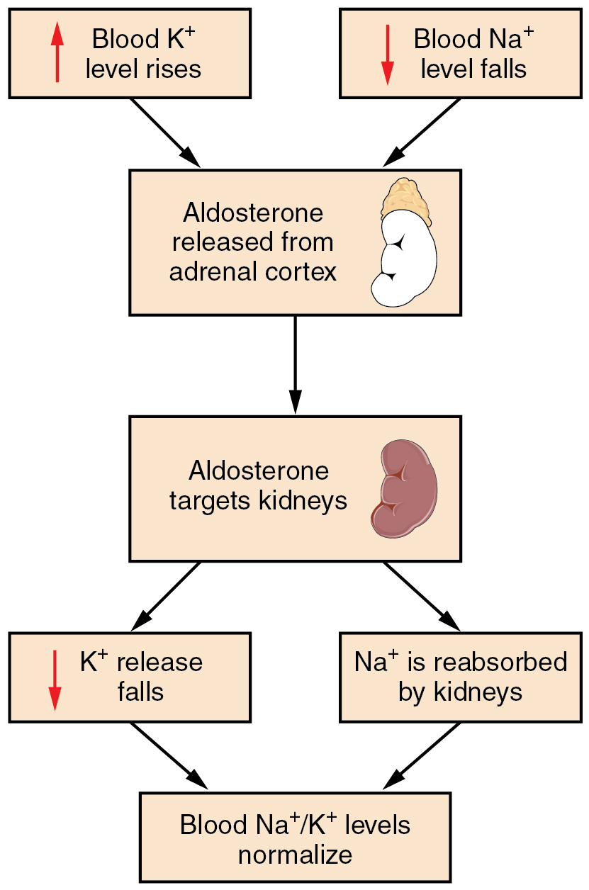
When aldosterone levels increase, Na+ is reabsorbed and more K+ is secreted (leading to excretion by the kidney); when aldosterone levels decrease, less Na+ is reabsorbed (leading to excretion by the kidney), more K+ is retained.
Aldosterone release from the adrenal cortex increases when angiotensin II levels increase, and/or when blood K+ levels increase (Figure 14.2.1). Note that increased blood K+ directly stimulates the adrenal cortex to release aldosterone; it does not stimulate RAAS.
Chloride
Chloride is the predominant extracellular anion. Chloride is a major contributor to the osmotic pressure gradient between the ICF and ECF, and plays an important role in maintaining proper hydration. Chloride functions to balance cations in the ECF, maintaining the electrical neutrality of this fluid. The paths of secretion and reabsorption of chloride ions in the renal system follow the paths of sodium ions.
Hypochloremia, or lower-than-normal blood chloride levels, can occur because of defective renal tubular absorption. Vomiting, diarrhea, and metabolic acidosis can also lead to hypochloremia.
Hyperchloremia, or higher-than-normal blood chloride levels, can occur due to dehydration, excessive intake of dietary salt (NaCl) or swallowing of sea water, aspirin intoxication, congestive heart failure, and the hereditary, chronic lung disease, cystic fibrosis. In people who have cystic fibrosis, chloride levels in sweat are two to five times those of normal levels, and analysis of sweat is often used in the diagnosis of the disease.
Regulation of Cl–
Chloride is important in acid–base balance in the extracellular space and has other functions, such as in the stomach, where it combines with hydrogen ions in the stomach lumen to form hydrochloric acid, aiding digestion. Its close association with Na+ in the extracellular environment makes it the dominant anion of this compartment, and its regulation closely mirrors that of Na+.
Calcium and Phosphate
About 99% of the calcium in your body is bound up in bone in the form of calcium phosphate salts, which provides hardness to the bone and serves as a mineral reserve for calcium and its salts for the rest of the tissues. A little more than one-half of blood calcium is bound to proteins, leaving the rest in its ionized form. Calcium ions, Ca2+, are necessary for muscle contraction, enzyme activity, and blood coagulation. In addition, Ca2+ helps to stabilize cell membranes and is essential for the release of neurotransmitters from neurons and of hormones from endocrine glands.
Calcium is absorbed through the intestines under the influence of activated vitamin D (calcitriol, or dihydroxyvitamin D). A deficiency of vitamin D leads to a decrease in absorbed calcium and, eventually, a depletion of calcium stores from the skeletal system, potentially leading to rickets in children and osteomalacia in adults, contributing to osteoporosis.
Hypocalcemia, or abnormally low calcium blood levels, is seen in hypoparathyroidism, which may follow the removal of the thyroid gland, because the four nodules of the parathyroid gland are embedded in it. This can lead to cardiac depression, increased neuromuscular excitability, muscular cramps, and skeletal weakness. Hypercalcemia, or abnormally high calcium blood levels, is seen in primary hyperparathyroidism. This can lead to cardiac arrhythmias and arrest, muscle weakness, CNS confusion, and coma. Some malignancies may also result in hypercalcemia.
Phosphate is present in the blood in two ionic forms: H2PO4 – and HPO42-. Bone binds up 85% of the body’s phosphate as part of calcium-phosphate salts. Phosphate is found in phospholipids, such as those that make up the cell membrane, and in ATP, nucleotides, and buffers.
Hypophosphatemia, or abnormally low phosphate blood levels, occurs with heavy use of antacids, during alcohol withdrawal, and during malnourishment. In the face of phosphate depletion, the kidneys usually conserve phosphate, but during starvation, this conservation is impaired greatly.
Hyperphosphatemia, or abnormally increased levels of phosphates in the blood, occurs if there is decreased renal function or in cases of acute lymphocytic leukemia. Additionally, because phosphate is a major constituent of the ICF, any significant destruction of cells can result in dumping of phosphate into the ECF.
Regulation of Calcium and Phosphate
Calcium and phosphate are both regulated through the actions of three hormones: parathyroid hormone (PTH), dihydroxyvitamin D (calcitriol), and calcitonin. All three are released or synthesized in response to the blood levels of calcium.
PTH is released from the parathyroid glands in response to a decrease in the concentration of blood Ca2+. PTH activates osteoclasts to break down bone matrix; this releases Ca2+ and HPO42- into the blood. PTH also increases the gastrointestinal absorption of dietary calcium by converting vitamin D into dihydroxyvitamin D (calcitriol), an active form of vitamin D that intestinal epithelial cells require to absorb calcium.
PTH also raises blood calcium levels by increasing calcium reabsorption by the DCT of the nephron. PTH also decreases the reabsorption of phosphate through the kidneys.
Calcitonin is released from the thyroid gland in response to elevated blood levels of calcium. The hormone increases the activity of osteoblasts, which remove calcium from the blood and incorporate calcium into the bony matrix. Calcitonin also decreases the reabsorption of phosphate by the nephron, lowering blood phosphate.
Bicarbonate
Bicarbonate is the second most abundant anion in the blood. Its principal function is to maintain the body’s acid-base balance by being part of buffer systems. This role will be discussed in the next section of this chapter.
Bicarbonate ions result from a chemical reaction that starts with carbon dioxide (CO2) and water, two molecules that are produced at the end of aerobic metabolism. Carbon dioxide combines with water to form to form carbonic acid (H2CO3), which dissociates into two ions: bicarbonate (HCO3–) and hydrogen (H+). The following formula depicts this reaction:
This reaction occurs spontaneously, but also is sped up by the action of the enzyme carbonic anhydrase (CA) that catalyzes the formation of carbonic acid from carbon dioxide and water.
The bidirectional arrows indicate that the reactions can proceed in either direction, depending on the concentrations of the reactants and products.
Regulation of Bicarbonate, H+, and pH
The acid–base homeostasis of the body is a function of chemical buffers and physiologic buffering provided by the lungs and kidneys. Buffers, especially proteins, HCO3−, and ammonia have a very large capacity to absorb or release H+ as needed to resist a change in pH. They can act within fractions of a second.
The respiratory system can alter plasma pH rapidly (seconds to minutes) by altering ventilation rate and changing plasma PCO2; this in turn will change plasma H+ by changing the rate of the forward or reverse reaction in the carbonic acid-bicarbonate buffer system:
- hyperventilation -> decreased plasma CO2 -> decreased plasma H+ -> increased plasma pH
- hypoventilation -> increased plasma CO2 -> increased plasma H+ -> decreased plasma pH
The kidneys can alter blood HCO3− and H+ to regulate blood pH. The renal capacity to maintain acid-base balance is large but slow (minutes to hours). The cells of the PCT and DCT actively secrete H+ into the forming urine via different mechanisms while simultaneously reabsorbing HCO3– . The detailed mechanism by which this occurs is discussed in the next section of this chapter.
Section Review
Electrolytes serve various purposes, such as helping to conduct electrical impulses along cell membranes in neurons and muscles, stabilizing enzyme structures, and releasing hormones from endocrine glands. The electrolytes in plasma also contribute to the osmotic balance that controls the movement of water between cells and their environment. Imbalances of these electrolytes can result in various problems in the body, and their concentrations are tightly regulated.
Sodium is the major cation of the ECF. It is the electrolyte that contributes the most to the osmotic pressure of the ECF and it plays a main role in determining ECF volume and body water distribution. Potassium is the main cation found in the ICF.
Aldosterone and angiotensin II control the exchange of sodium and potassium between the renal filtrate and the renal collecting tubule. Calcium and phosphate are regulated by PTH, calcitrol, and calcitonin.
Hormonal control of fluid volume occurs primarily via the renin-angiotensin-aldosterone system (RAAS) and atrial natriuretic peptide (ANP). Progesterone is similar in structure to aldosterone and can bind to and weakly stimulate aldosterone receptors, providing a similar but diminished response.
Blood pressure is a reflection of blood volume and is sensed by baroreceptors in the aortic arch and carotid sinuses, granular cells in the juxtaglomerular apparatus, and cardiac muscle fibers in the atria.
Acid-base balance (blood pH regulation) occurs through chemical buffers and the activities of the respiratory and renal systems. The respiratory system maintains acid-base balance by changing plasma PCO2 , and the kidneys alter reabsorption of HCO3– and secretion of H+ in order to stabilize plasma pH.
Review Questions
Critical Thinking Questions
Glossary
- ANP (atrial natriuretic peptide)
- hormone produced by the atria; decreases the reabsorption of sodium by nephrons
- calcitriol
- active form of vitamin D required by the intestinal epithelial cells for the absorption of calcium; also called dihydroxyvitamin D
- carbonic anhydrase
- enzyme that catalyzes the formation of carbonic acid from carbon dioxide and water
- hypercalcemia
- abnormally increased blood levels of calcium
- hyperchloremia
- higher-than-normal blood chloride levels
- hyperkalemia
- higher-than-normal blood potassium levels
- hypernatremia
- abnormal increase in blood sodium levels
- hyperphosphatemia
- abnormally increased blood phosphate levels
- hypocalcemia
- abnormally low blood levels of calcium
- hypochloremia
- lower-than-normal blood chloride levels
- hypokalemia
- abnormally decreased blood levels of potassium
- hyponatremia
- lower-than-normal levels of sodium in the blood
- hypophosphatemia
- abnormally low blood phosphate levels
Glossary Flashcards
This work, Human Physiology, is adapted from Anatomy & Physiology by OpenStax, licensed under CC BY. This edition, with revised content and artwork, is licensed under CC BY-SA except where otherwise noted.
Images from Anatomy & Physiology by OpenStax are licensed under CC BY except where otherwise noted.
Access the original for free at OpenStax.
Report an Error
Did you find an error, typo, broken link, or other problem in the text? Please follow this link to the error reporting form to submit an error report to the authors.
