Chapter 18. The Sexual Systems
18.2 Development of Sexual Anatomy
Learning Objectives
By the end of this section, you will be able to:
- describe the role of chromosomes (X and Y) and the SRY in sex determination;
- compare and contrast the actions of androgens and estrogens on the development of the gonads during fetal development;
- identify and explain two different mechanisms by which intersex traits can arise;
- define puberty;
- describe changes in the hypothalamic-pituitary-gonadal axis that occur at the onset of puberty;
- identify, compare, and contrast the development of secondary sex characteristics in prototypical males and females;
- list the major targets of estrogens and progesterone and explain the target responses (e.g., oogenesis, breast development, fat deposition); and
- list the major target organs and tissues of testosterone and related androgens and explain the hormonal mechanisms of action and the target responses (e.g., sperm production, fat deposition, muscle development).
Introduction
The development of the sexual systems begins soon after fertilization of the egg, with primordial gonads beginning to develop approximately one month after conception. Sexual system development continues in utero, but there is little change in the system between infancy and puberty.
Development of the Sexual Organs in the Embryo and Fetus
The development of both sex organs is an active process. It used be thought that without chemical prompting, all fertilized eggs would develop a clitoris and vagina. However, without androgenic chemical signaling initiated by the SRY (sex-determining region of the Y chromosome) gene on the Y chromosome, an embryo will not develop a penis and scrotum. Individuals without a Y chromosome also do not have the SRY gene. Without a functional SRY gene, an individual will typically develop a uterus and ovaries if crucial genes are also present.
In all embryos, the same group of cells has the potential to develop into either testes and ovaries; this tissue is considered bipotential. The SRY gene actively recruits other genes that begin to develop the testes, and suppresses other genes that would lead to development of ovaries. As part of this SRY-prompted cascade, germ cells in the bipotential gonads differentiate into spermatogonia. Without SRY, different genes are expressed, oogonia form, and primordial follicles develop in the primitive ovary.
Soon after the formation of the testis, the interstitial (Leydig) cells begin to secrete testosterone. Testosterone can influence tissues that are bipotential. For example, with exposure to testosterone, cells that could become either the glans penis or the glans clitoris form the glans penis. Without testosterone, these same cells differentiate into the clitoris.
Not all tissues in the reproductive tract are bipotential. The internal reproductive structures (for example the uterus, uterine tubes, and part of the vagina; and the epididymis, ductus deferens, and seminal vesicles) form from one of two rudimentary duct systems in the embryo.
Development of the internal sexual organs requires one set of ducts to develop and the other set to degrade. A hormone secreted from sustentacular (Sertoli) cells trigger a degradation of the paramesonephric (Müllerian) duct, and therefore a uterus is unlikely to develop. At the same time, testosterone secretion stimulates growth of the mesonephric (Wolffian) duct, leading to development of the epididymis and vas deferens. Without such sustentacular cell hormone secretion, the paramesonephric duct will now develop; and without testosterone, the mesonephric duct will degrade. Thus, the offspring in this circumstance will likely develop a uterus, and not an epididymis or vas deferens. For more information and a figure of differentiation of the gonads, seek additional content on fetal development.
There are many reasons why sexual anatomy would develop differently than previously described, and it is important to locate intersex anatomy on the spectrum of normal human variation between the binary female and male. In some cases, the receptors that the hormones typically bind to do not develop. For example, in the case of androgen insensitivity, an individual with XY chromosomes, and an SRY gene, will still produce hormones from the sustentacular cells that lead to degradation of the paramesonephric duct—meaning that no uterus can develop. They will also develop testes which will produce testosterone, (androgens) but the cells can not react to the hormones because they lack the receptor to bind the hormone. Therefore, the epididymis and vas deferens are not produced, and the external genitalia develop into a clitoris and vagina. The result is an individual with XY chromosomes, non-descended testes, clitoris and vagina but no uterus.
In contrast to the example above, an intersex condition can result from having hormone secretion beyond what is expected based on the chromosomes. In Congenital Adrenal Hyperplasia, individuals with XX chromosomes have an increase in androgens produced by adrenal glands. The result is a clitoris that is enlarged in size, and at birth may appear similar to a penis. The following image illustrates the spectrum that can exist in clitoral size during adrenal hyperplasia. The increased androgen production in these XX individuals may also lead to increased body hair, receding hair line, deep voice and muscular physique. In an XY individual, a decrease in the expected androgen production can lead to a penis that is much smaller than average, and termed micropenis. This reinforces the notion that external genitalia are developed across a spectrum of size between a clitoris and penis based on the degree of exposure to androgens. This spectrum of normal human variation does not require surgical treatment, only an open mind to the notion of what normal variation might include. Individuals with intersex anatomy have no additional health risks when left to develop on their own, while surgical intervention at a young age includes the risk of surgical complications including nerve damage and infection.
Onset of Puberty
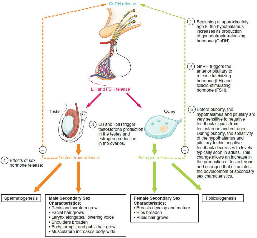
Puberty is the stage of development at which individuals become sexually mature. As shown in Figure 18.2.1, a concerted release of hormones from the hypothalamus (GnRH), the anterior pituitary (LH and FSH), and the gonads (either testosterone or estrogen) is responsible for the maturation of the reproductive systems and the development of secondary sex characteristics, which are physical changes in the body.
The first changes begin around the age of eight or nine when the production of LH becomes detectable. The release of LH occurs primarily at night during sleep and precedes the physical changes of puberty by several years. In pre-pubescent children, the sensitivity of the negative feedback system in the hypothalamus and pituitary is very high. This means that very low concentrations of androgens or estrogens will negatively feedback onto the hypothalamus and pituitary, keeping the production of GnRH, LH, and FSH low.
As an individual approaches puberty, two changes in sensitivity occur. The first is a decrease of sensitivity in the hypothalamus and pituitary to negative feedback, meaning that it takes increasingly larger concentrations of sex steroid hormones to stop the production of LH and FSH. The second change in sensitivity is an increase in sensitivity of the gonads to the FSH and LH signals, meaning the gonads of adults are more responsive to gonadotropins than are the gonads of children. Because of these two changes, the levels of LH and FSH slowly increase and lead to the enlargement and maturation of the gonads, which in turn leads to secretion of higher levels of sex hormones and the initiation of spermatogenesis and folliculogenesis.
In addition to age, multiple factors can affect the age of onset of puberty, including genetics, environment, and psychological stress. One of the more important influences may be nutrition; historical data demonstrate the effect of better and more consistent nutrition on the age of menarche in the United States, which decreased from an average age of approximately 17 years of age in 1860 to the current age of approximately 12.75 years in 1960, as it remains today. Some studies indicate a link between puberty onset and the amount of stored fat in an individual. This effect has been documented in both sexes. Body fat, corresponding with secretion of the hormone leptin by adipose cells, appears to have a strong role in determining menarche. This may reflect to some extent the high metabolic costs of gestation and lactation. In individuals who are lean and highly active, such as gymnasts, there is often a delay in the onset of puberty.
Signs of Puberty
Different sex steroid hormone concentrations also contribute to the development and function of secondary sexual characteristics. Examples of secondary sexual characteristics due to a predominance of testosterone or estrogen are listed in Table 18.1.
| Testosterone | Estrogen |
|---|---|
| Increased larynx size and deepening of the voice | Deposition of fat, predominantly in breasts and hips |
| Increased muscular development | Breast development |
| Growth of facial, axillary, and pubic hair, and increased growth of body hair | Broadening of the pelvis and growth of axillary and pubic hair |
An increased production of estrogen at puberty typically leads to the development of breast tissue. This is followed by the growth of axillary and pubic hair. A growth spurt typically starts at approximately age 9 to 11 and may last two years or more. During this time, an individual’s height can increase an average of 3 inches a year. The next step in puberty due to estrogen is menarche, the start of menstruation (also known as menses), which is the shedding of inner portion of the endometrium.
An increased production of testosterone leads to growth of the testes, typically the first physical sign of the beginning of puberty, which is followed by growth and pigmentation of the scrotum and growth of the penis. The next step is the growth of hair, including armpit, pubic, chest, and facial hair. Testosterone stimulates the growth of the larynx and thickening and lengthening of the vocal folds, which causes the voice to drop in pitch. The first fertile ejaculations typically appear at approximately 15 years of age, but this age can vary widely across individuals. The prostate normally doubles in size during puberty. A growth spurt occurs toward the end of puberty, at approximately age 11 to 13, and height can increase as much as 4 inches a year. In some individuals, pubertal development can continue through the early twenties.
Section Review
The reproductive systems of males and females begin to develop soon after conception. A gene on the Y chromosome called SRY is critical in stimulating a cascade of events that simultaneously stimulate testis development and repress the development of female structures. Testosterone produced by Leydig cells in the embryonic testis stimulates the development of male sexual organs. If testosterone is not present, female sexual organs will develop.
Whereas the gonads and some other reproductive tissues are considered bipotential, the tissue that forms the internal reproductive structures stems from ducts that will develop into only male (Wolffian) or female (Müllerian) structures. To be able to reproduce as an adult, one of these systems must develop properly and the other must degrade.
Further development of the reproductive systems occurs at puberty. The initiation of the changes that occur in puberty is the result of a decrease in sensitivity to negative feedback in the hypothalamus and pituitary gland, and an increase in sensitivity of the gonads to FSH and LH stimulation. These changes lead to increases in either estrogen or testosterone. The increase in sex steroid hormones leads to maturation of the gonads and other reproductive organs. The initiation of spermatogenesis begins, as well as ovulation and menstruation. Increases in sex steroid hormones also lead to the development of secondary sex characteristics.
Review Questions
Critical Thinking Questions
Glossary
- gonadotropin-releasing hormone (GnRH)
- hormone released by the hypothalamus that regulates the production of follicle-stimulating hormone and luteinizing hormone from the pituitary gland
- menarche
- first menstruation in a pubertal female
- menses
- shedding of the inner portion of the endometrium out though the vagina; also referred to as menstruation
- Müllerian duct
- duct system present in the embryo that will eventually form the internal female reproductive structures
- puberty
- life stage during which a male or female adolescent becomes anatomically and physiologically capable of reproduction
- secondary sex characteristics
- physical characteristics that are influenced by sex steroid hormones and have supporting roles in reproductive function
- Wolffian duct
- duct system present in the embryo that will eventually form the internal male reproductive structures
Glossary Flashcards
This work, Human Physiology, is adapted from Anatomy & Physiology by OpenStax, licensed under CC BY. This edition, with revised content and artwork, is licensed under CC BY-SA except where otherwise noted.
Images from Anatomy & Physiology by OpenStax are licensed under CC BY except where otherwise noted.
Access the original for free at OpenStax.
Report an Error
Did you find an error, typo, broken link, or other problem in the text? Please follow this link to the error reporting form to submit an error report to the authors.
