Chapter 5. The Nervous System and Nervous Tissue
5.1 Structure and Function of the Nervous System
Learning Objectives
By the end of this section, you will be able to:
- Identify the branches of the nervous system and their relevant sub-branches
- List the basic functions of the nervous system
The Central and Peripheral Nervous Systems
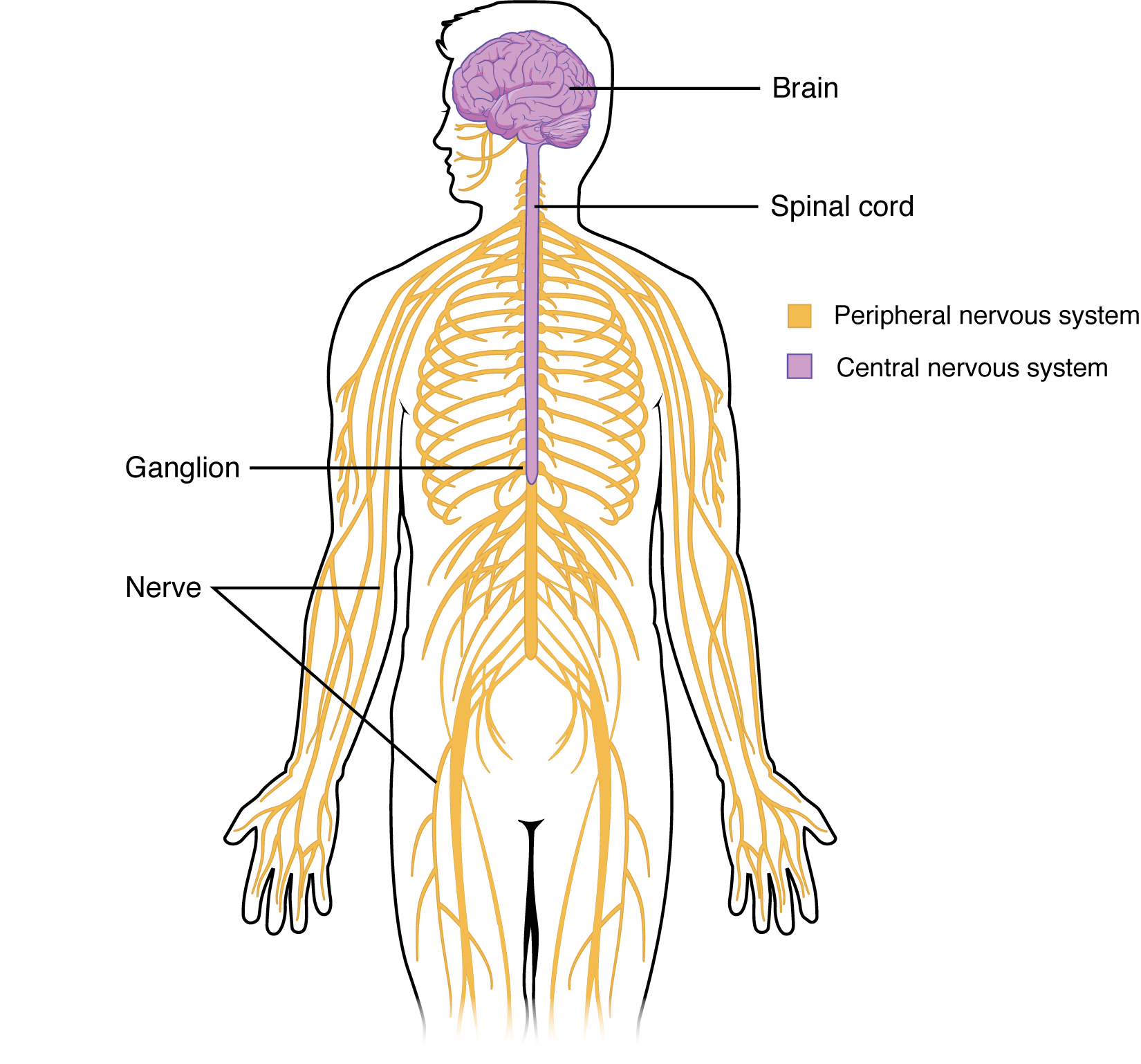
The picture you have in your mind of the nervous system probably includes the brain, the nervous tissue contained within the cranium, and the spinal cord, the extension of nervous tissue within the vertebral column. Additionally, the nervous tissue that reach out from the brain and spinal cord to the rest of the body (nerves) are also part of the nervous system. We can anatomically divide the nervous system into two major regions: the central nervous system (CNS) is the brain and spinal cord, and the peripheral nervous system (PNS) is the nerves (Figure 5.1.1). The brain is contained within the cranial cavity of the skull, and the spinal cord is contained within the vertebral canal of the vertebral column. The peripheral nervous system is so named because it is in the periphery—meaning beyond the brain and spinal cord. The peripheral nervous system can be further divided into efferent (motor) branch and the afferent (sensory) branch. The efferent branch consists of the somatic nervous system and autonomic nervous system. Some scholars also include the visceral nervous system as a branch of the peripheral nervous system. The peripheral nervous system will be discussed in later chapters.
Functional Divisions of the Nervous System
In addition to the anatomical divisions listed above, the nervous system can also be divided on the basis of its functions. The nervous system is involved in receiving information about the environment around us (sensory functions, sensation), generating responses to that information (motor functions, response), and coordinating the two (integration).
Sensation. Sensation refers to receiving information about the environment, either what is happening outside (i.e., heat from the sun) or inside the body (i.e., heat from muscle activity). These sensations are known as stimuli (singular stimulus), and different sensory receptors are responsible for detecting different stimuli. Sensory information travels towards the CNS through the PNS nerves in the specific division known as the afferent (sensory) branch of the PNS. When information arises from sensory receptors in the skin, skeletal muscles, or joints, it is transmitted to the CNS using somatic sensory neurons; when information arises from sensory receptors in the blood vessels or internal organs, it is transmitted to the CNS using visceral sensory neurons.
Response. The nervous system produces a response in effector organs (such as muscles or glands) due to the sensory stimuli. The motor (efferent) branch of the PNS carries signals away from the CNS to the effector organs. When the effector organ is a skeletal muscle, the neuron carrying the information is called a somatic motor neuron; when the effector organ is cardiac or smooth muscle or glandular tissue, the neuron carrying the information is called an autonomic motor neuron. Voluntary responses are governed by somatic motor neurons and involuntary responses are governed by the autonomic motor neurons, which are discussed in the next section.
Integration. Stimuli that are detected by sensory structures are communicated to the nervous system where information is processed. In the CNS, information from some stimuli is compared with, or integrated with, information from other stimuli or memories of previous stimuli. Then, a motor neuron is activated to initiate a response from the effector organ. This process during which sensory information is processed and a motor response generated is called integration (Figure 5.1.2).
Section Review
The nervous system can be separated into divisions on the basis of anatomy and physiology. The anatomical divisions are the central and peripheral nervous systems. The CNS is the brain and spinal cord. The PNS is everything else and includes afferent and efferent branches with further subdivisions for somatic, visceral, and autonomic function. Functionally, the nervous system can be divided into those regions that are responsible for sensation, those that are responsible for integration, and those that are responsible for generating responses.
Review Questions
Critical Thinking Questions
Glossary
- autonomic nervous system
- functional division of the efferent branch of the PNS that is responsible for control of cardiac and smooth muscle, as well as glandular tissue
- brain
- the large organ of the central nervous system contained within the cranium and continuous with the spinal cord
- central nervous system (CNS)
- anatomical division of the nervous system that includes the brain and spinal cord
- integration
- nervous system function that processes sensory perceptions and produce a response
- peripheral nervous system (PNS)
- anatomical division of the nervous system that extends from the brain and spinal cord to the rest of the body
- response
- nervous system function that causes a target tissue (muscle or gland) to produce an event as a consequence to stimuli
- sensation
- nervous system function that receives information from the environment and translates it into the electrical signals of nervous tissue
- somatic nervous system
- functional division of the nervous system that is concerned with conscious perception, voluntary movement, and skeletal muscle reflexes
- spinal cord
- organ of the central nervous system found within the vertebral cavity and connected with the periphery through spinal nerves; mediates reflex behaviors
- stimulus
- an event in the external or internal environment that registers as activity in a sensory neuron
Glossary Flashcards
This work, Human Physiology, is adapted from Anatomy & Physiology by OpenStax, licensed under CC BY. This edition, with revised content and artwork, is licensed under CC BY-SA except where otherwise noted.
Images from Anatomy & Physiology by OpenStax are licensed under CC BY except where otherwise noted.
Access the original for free at OpenStax.
Image Descriptions
Figure 5.1.1. This anatomical diagram shows the human nervous system within an outlined human figure viewed from the side (head) and front (body). The diagram uses color-coding to distinguish the central nervous system (purple) from the peripheral nervous system (yellow/orange). The purple brain is shown at the top with visible surface texture, connected to a purple spinal cord that runs down through the neck and back. Yellow/orange nerve pathways branch symmetrically from the spinal cord throughout the torso, arms, and legs in a tree-like pattern. Labels identify the brain, spinal cord, ganglion, and individual nerves, with a legend indicating the color coding for central and peripheral nervous systems. [Return to Figure 5.1.1]
Figure 5.1.2. This diagram illustrates the communication pathway between the peripheral and central nervous systems, divided into two color-coded sections: green (left) for the peripheral nervous system and blue (right) for the central nervous system. The left side shows sensory information collection, featuring an illustrated eye at the top and a red muscle (effector organ) at the bottom, both connected by orange nerve pathways. The right side displays a purple brain connected to a spinal cord, labeled as the integration center. Orange afferent neurons carry signals from the eye toward the brain (shown by a black arrow pointing right), where the brain processes the information. Orange efferent neurons then transmit signals back from the spinal cord to the muscle (shown by a black arrow pointing left), demonstrating the complete sensory-motor reflex arc. Small purple dots along the neural pathways represent neuron cell bodies, illustrating how the nervous system receives sensory input, processes it centrally, and produces a motor response. [Return to Figure 5.1.2]
Report an Error
Did you find an error, typo, broken link, or other problem in the text? Please follow this link to the error reporting form to submit an error report to the authors.
