Chapter 5. The Nervous System and Nervous Tissue
5.2 Nervous Tissue
Learning Objectives
By the end of this section, you will be able to:
- Name the cells of the nervous system and cite their function
- Outline the parts of the neuron and relate it to their functional role
- List the glial cells of the CNS and PNS and describe their function
- Describe the role of neuroglia in increasing conduction velocity via myelination of neurons
- Analyze how demyelinating diseases disproportionately impact Black patients and possible ways to advance care and outcomes for these patients
Nervous tissue is composed of two types of cells: neurons and glial cells. Neurons are responsible for the computation and communication that the nervous system provides. They are electrically active and release chemical signals to communicate between each other and with target cells. Glial cells, or glia or neuroglia, are much smaller than neurons and play a supporting role for nervous tissue. Glial cells maintain the extracellular environment around neurons, improve signal conduction in neurons, and protect them from pathogens. Ongoing research also suggests that glial cell number matches neuron number and that they even can send signals themselves.
Neuron Anatomy
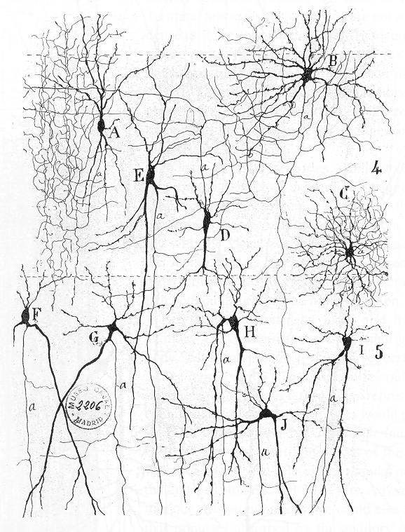
Neurons are nucleated cells with specialized structural properties. Some neurons have a single long extension (axon) that reaches great distances, while others are very small, star-shaped cells without obvious axons (Figure 5.2.1).
Though neuron shapes vary greatly, every neuron houses its nucleus in a region known as the soma (also called the cell body) from which cellular activity like repair or cell membrane recycling is controlled. The soma can also receive signals from other neurons. Associated with the nucleus, neurons also have many rough endoplasmic reticula called Nissl bodies (which can be seen in neurons using a light microscope). The nucleus, Nissl bodies, and Golgi apparatuses together produce the many ion channels and pumps that reside in the cell membrane. These transmembrane proteins are necessary for neurons to send electrical signals (graded potentials and action potentials; see Section 5.4). In addition, neurons consume much ATP and typically have many mitochondria.
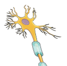
In Figure 5.2.2, the cell body shows both many short projections and one long projection emerging from the cell body. These short projections are dendrites, which receive most of the input from other neurons or stimuli in the extracellular environment; the location of the dendrites on the neuron marks the receptive region of the neuron. Dendrites are usually highly branched processes, providing locations for other neurons to communicate with the neuron. Neurons have polarity—meaning that information flows in one direction through the neuron. In the neuron in Figure 5.2.2, information flows from the dendrites, across the cell body, and down the large axon emerging from the cell body at the axon hillock (an anatomical term to describe where the cell body and axon meet). The first section of the axon where an action potential is generated is called the initial segment. In multipolar and bipolar neurons, the initial segment is found at the axon hillock (Figure 5.2.3). However, in unipolar neurons, the initial segment is not found at the axon hillock and can actually be located many inches or even a few feet from it near the dendrites (Figure 5.2.3)! Often axons are wrapped by myelin sheaths, leaving exposed sections (node of Ranvier) between segments of myelin. Myelin is produced by oligodendrocytes (glial cells) in the CNS and Schwann cells in the PNS; it acts as electrical insulation, speeding information conduction down the neuron. Once information reaches the axon terminal (terminal end) of this neuron, it is transferred to another cell. The site of communication between a neuron and its target cell is called a synapse. The terminal end has several branches, each with a synaptic end bulb to store chemicals needed for communication with the next cell. Figure 5.2.2 shows the relationship of these parts to one another.
Types of Neurons
There are trillions of neurons in the nervous system, and cell shape can vary widely. Three common shapes of neurons are shown in Figure 5.2.3.
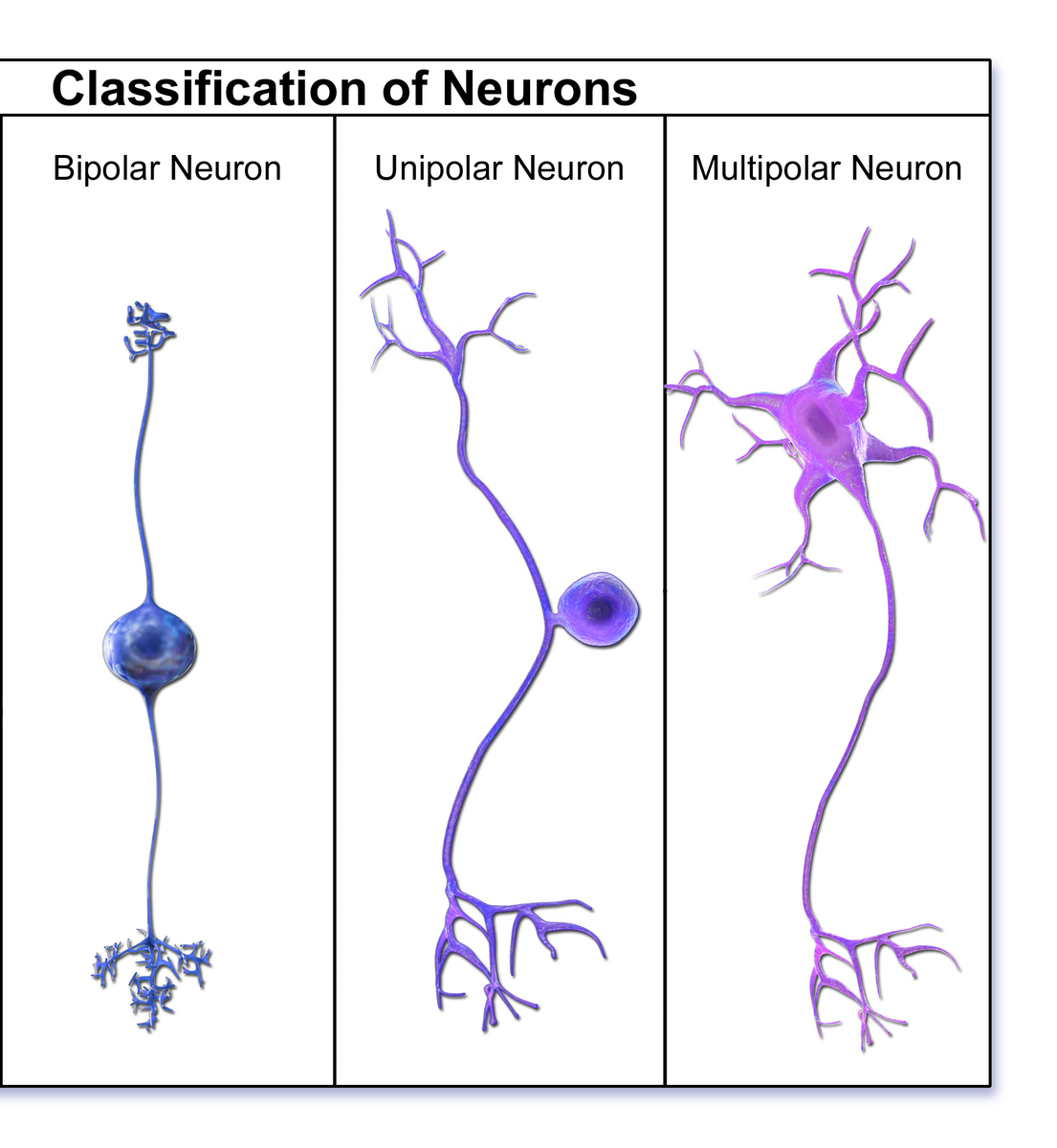
Multipolar neurons have multiple processes emerging from their cell bodies (hence the name multipolar). They have dendrites attached to their cell bodies and often one long axon. Motor neurons are multipolar neurons, as are many neurons of the CNS.
Bipolar neurons have two processes, which extend from each end of the cell body opposite to each other. One is the axon and one the dendrite. Bipolar neurons are not very common. They are found mainly in the olfactory epithelium (where smell stimuli are sensed) and as part of the retina in the eye.
Unipolar neurons have one long axon emerging from the cell body, with the cell body located between the two ends and off to the side. At one end of the axon are dendrites, and at the other end, the axon forms synaptic connections with a target cell. Unipolar neurons are exclusively sensory neurons and have their dendrites in the periphery where they detect stimuli. Their cell bodies are typically found in ganglia of the PNS.
Glial Cells
There are six types of glial cells. Four of them are found in the CNS and two are found in the PNS. Table 5.2 outlines some common characteristics and functions.
| CNS glia image |  |
 |
 |
 |
|---|---|---|---|---|
| CNS glia name | Astrocytes | Oligodendrocyte | Microglia | Ependymal cell |
| PNS glia image |  |
 |
– | – |
| PNS glia name | Satellite cell | Schwann Cell | – | – |
| Functions | Maintain extracellular environment
Remove excess neurotransmitter Direct neural growth Induce blood-brain barrier in CNS (astrocyte only) |
Create myelin | Immune surveillance and phagocytosis | Create and circulate Cerebrospinal fluid (CSF) |
Glial Cells of the CNS
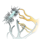
One cell providing support to neurons of the CNS is the astrocyte, so named because it appears to be star-shaped under the microscope (“astro-” meaning “star” and “-cyte” meaning “cell”). Astrocytes have many processes extending from their main cell body (not axons or dendrites like neurons, just cell extensions). Those processes extend to interact with neurons, blood vessels, or the connective tissue covering the CNS. Generally, they are supporting cells for the neurons in the CNS. Some ways in which they support neurons in the CNS are by maintaining the concentration of chemicals in the extracellular space, removing excess signaling molecules, reacting to tissue damage, and inducing to the blood-brain barrier (BBB). The BBB is a protective physiological barrier that keeps many substances that circulate in the blood from getting into the CNS, restricting what can cross from circulating blood into the CNS. Usually, blood vessels are leaky because there are gaps between the cells of the vessel walls. These gaps permit rapid movement of molecules out of the blood into the extracellular space around tissue cells, delivering nutrients and hormones. However, the neurons of the brain may be affected by rapid, regular changes in extracellular concentrations preventing signal transmission. To prevent such fluctuations, astrocytes release compounds to the blood vessels, inducing tight junctions between the otherwise leaky blood vessel cells. When the BBB is intact, nutrient molecules, such as glucose or amino acids, must now pass through the vessel cells of the BBB by transcellular processes (using membrane proteins). Small, fat-soluble molecules (respiratory gases, alcohol) are able simply diffuse through the cell membranes, but other large, water-soluble molecules cannot. The highly restrictive permeability of the BBB may restrict drug delivery to the CNS. Pharmaceutical companies are challenged to design drugs that can cross the BBB as well as have an effect on the nervous system.
Also found in CNS tissue is the oligodendrocyte, sometimes called just “oligo,” which is the glial cell type that insulates axons in the CNS. The name means “cell of a few branches” (“oligo-” meaning “few”; “dendro-” meaning “branches”). There are a few processes that extend from the cell body. Each one reaches out and surrounds an axon to insulate it in myelin. One oligodendrocyte will provide the myelin for multiple axon segments, either for the same axon or for separate axons. The function of myelin will be discussed below.
Microglia are, as the name implies, smaller than most of the other glial cells. Ongoing research into these cells, although not entirely conclusive, suggests that they may originate as white blood cells, called macrophages, that become part of the CNS during early development. While their origin is not conclusively determined, their function is related to what macrophages do in the rest of the body. When macrophages encounter diseased or damaged cells in the rest of the body, they ingest and digest those cells or the pathogens that cause disease. Microglia are the cells in the CNS that can do this in normal, healthy tissue, and they are therefore also referred to as CNS-resident macrophages.
Ependymal cells filter blood to make cerebrospinal fluid (CSF), the fluid that circulates through the CNS. CSF is needed in the brain to provide nutrients, remove wastes and create a stable extracellular environment because the BBB is so restrictive. In each of the brain cavities (ventricles), ependymal cells surround the blood vessels forming choroid plexuses. These choroid plexuses filter specific components of the blood to produce cerebrospinal fluid. Every day they produce enough CSF to fill a pint glass! Though the BBB is absent in the choroid plexuses, the ependymal cells there are connected to each other by tight connections, forming a highly restrictive boundary. More ependymal cells line the ventricles and use their cilia to help move the CSF through the ventricular space. The relationship of these glial cells to the structure of the CNS is seen in Table 5.2.
Glial Cells of the PNS
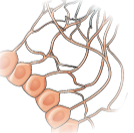
One of the two types of glial cells found in the PNS is the satellite cell. Satellite cells surround the cell bodies of neurons in the PNS. They provide support, performing similar functions in the periphery as astrocytes do in the CNS—except, of course, for establishing the BBB.
The second type of glial cell is the Schwann cell, which insulate axons with myelin in the periphery. Schwann cells are different than oligodendrocytes in that a Schwann cell wraps around a portion of only one axon segment and no others. Oligodendrocytes have processes that reach out to multiple axon segments, whereas the entire Schwann cell surrounds just one axon segment. The nucleus and cytoplasm of the Schwann cell are on the edge of the myelin sheath. The relationship of these two types of glial cells to ganglia and nerves in the PNS is seen in Figure 5.2.4.
Myelin
Oligodendrocytes in the CNS and Schwann cells in the PNS provide myelin. While the manner in which either cell is associated with the axon segment, or segments, that it insulates is different, the means of myelinating an axon segment is mostly the same in the two situations. Myelin is a lipid-rich sheath that surrounds the axon and by doing so creates a myelin sheath that facilitates the transmission of electrical signals along the axon. Simply, myelinated axons send signals faster than unmyelinated axons. The lipids of myelin are essentially the phospholipids of the glial cell membrane. Myelin, however, is more than just the membrane of the glial cell. It also includes important proteins that are integral to that membrane. Some of the proteins help to hold the layers of the glial cell membrane closely together.
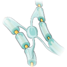
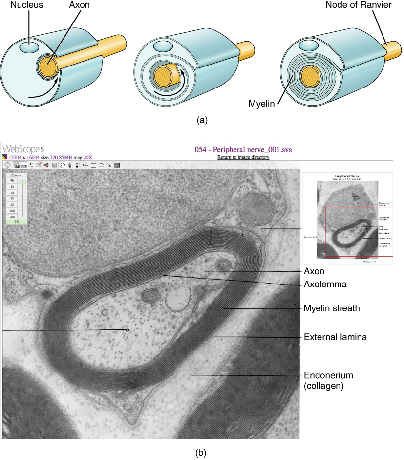
The appearance of the myelin sheath can be thought of as similar to the pastry wrapped around a hot dog for “pigs in a blanket.” The glial cell is wrapped around the axon several times with little to no cytoplasm between the glial cell layers. For oligodendrocytes, the rest of the cell is separate from the myelin sheath as a cell process extends back toward the cell body. A few other processes provide the same insulation for other axon segments in the area. For Schwann cells, the outermost layer of the cell membrane contains cytoplasm and the nucleus of the cell as a bulge on one side of the myelin sheath. During development, the glial cell is loosely or incompletely wrapped around the axon (Figure 5.2.5a). The edges of this loose enclosure extend toward each other, and one end tucks under the other. The inner edge wraps around the axon, creating several layers, and the other edge closes around the outside so that the axon is completely enclosed.
External Website

View the University of Michigan WebScope’s website to see an electron micrograph of a cross-section of a myelinated nerve fiber. The axon contains microtubules and neurofilaments bounded by a plasma membrane known as the axolemma. Outside the plasma membrane of the axon is the myelin sheath, which is composed of the tightly wrapped plasma membrane of a Schwann cell.
Myelin sheaths can extend for one or two millimeters, depending on the diameter of the axon. Axon diameters can be as small as 1 to 20 micrometers. Because a micrometer is 1/1000 of a millimeter, this means that the length of a myelin sheath can be 100 to 1,000 times the diameter of the axon. Figure 5.2.2, Figure 5.2.4, and Figure 5.2.5 show the myelin sheath surrounding an axon segment but are not to scale. If the myelin sheath were drawn to scale, the neuron would have to be immense—possibly covering an entire wall of the room in which you are sitting.
Demyelinating Diseases
Several diseases can result from the demyelination of axons. The causes of these diseases are not the same; some have genetic causes, some are caused by pathogens, and others are the result of autoimmune disorders. Though the causes are varied, the results are largely similar. The myelin insulation of axons is compromised, making electrical signaling slower. In some cases, signaling stops, preventing muscles from responding and causing paralysis.
Multiple sclerosis (MS) is one such disease. It is an example of an autoimmune disease. The antibodies produced by lymphocytes (a type of white blood cell) mark myelin as something that should not be in the body. This causes inflammation and the destruction of the myelin in the CNS. As the insulation around the axons is destroyed by the disease, scarring occurs. This is where the name of the disease comes from: sclerosis means hardening of tissue, as occurs in a scar. Multiple scars are found in the white matter of the brain and spinal cord. Control of the skeletal and smooth musculature is compromised, affecting not only movement but also control of organs such as the bladder.
Guillain-Barré (pronounced gee-YAN bah-RAY) syndrome is an example of a demyelinating disease of the PNS. It is also the result of an autoimmune reaction, but the inflammation is in peripheral nerves. Sensory symptoms or motor deficits are common, and autonomic failures can lead to changes in the heart rhythm or a drop in blood pressure, especially when standing, which causes dizziness.
Racial Health Inequities and Disparities in Demyelinating Diseases
Historically, research on demyelinating diseases has focused on White patients. Currently, Black and Hispanic populations in the United States are underrepresented in MS-related clinical trials. This underrepresentation contributes to our lack of knowledge on how to best treat and care for this population and how to advance long-term outcomes.
MS most commonly affects young women, primarily of northern European ancestry. However, newer studies have shown that Black patients have a higher MS risk and incidence. Furthermore, studies show that among people age 55 or less with MS, Black people have the highest mortality rate. Studies have shown that genetics, the environment, and immunology can be risk factors that contribute the pathogenesis of the disease. However, these risk factors do not account for the majority of MS risk. Social determinants of health, including income and education, can also influence MS and likely play a huge role.
Several social determinants can influence inequities in MS care and outcomes including: underrepresentation in clinical studies, systemic biases and racism, limited access to resources, lack of culturally competent care, systemic barriers to health care, socioeconomic status, and health literacy.
Due to the lack of research, it is difficult to determine the unmet needs of the Black population with MS. Recently, the National African Americans with MS Registry was developed to help address this lack of knowledge. They state: “The goals of NAAMS Registry are to have an accurate understanding of the number of African Americans afflicted with MS, their potential barriers to care, how this relates to where the patients live, and whether certain medications are of more benefit to African Americans.”
Section Review
Nervous tissue contains two major cell types: neurons and glial cells. Neurons are responsible for communication through electrical signals. Glial cells are supporting cells, allowing neuron function.
Though neuron shape varies, neurons are polarized cells, based on the flow of electrical signals along their membrane. In multipolar neurons, dendrites receive signals and pass them to the cell body; signals then propagate along the axon towards the terminal end that synapses with a target cell. Myelin on axons speeds signal conduction and is provided by different glial cells in the CNS and PNS.
The nervous system has several types of glial cells, categorized by the anatomical division in which they are found. In the CNS, astrocytes, oligodendrocytes, microglia, and ependymal cells perform different functions that support neurons. Astrocytes maintain the chemical environment around neurons and are crucial for regulating the BBB. Oligodendrocytes myelinate neurons, and microglia act as phagocytes and play a role in immune surveillance. Ependymal cells filter blood to produce CSF. CSF circulates through the CNS providing nutrients and removing waste. In the PNS, satellite cells maintain the extracellular environment around cell bodies, and Schwann cells insulate peripheral axons.
Review Questions
Critical Thinking Questions
Glossary
- astrocyte
- glial cell type of the CNS that provides support for neurons and maintains the BBB
- axon
- single process of the neuron that carries an electrical signal (action potential) away from the cell body toward a target cell
- axon hillock
- region of the neuron cell body that gives rise to the axon
- axon terminal (terminal end)
- end of the axon, where there are usually several branches extending toward the target cell
- bipolar
- shape of a neuron with two processes extending from the neuron cell body—the axon and one dendrite
- blood-brain barrier (BBB)
- physiological barrier between the circulatory system and the CNS that establishes a privileged blood supply, restricting the flow of substances into the CNS
- cerebrospinal fluid (CSF)
- circulatory medium within the CNS that is produced by ependymal cells in the choroid plexus filtering the blood
- choroid plexus
- specialized structure containing ependymal cells that line blood capillaries and filter blood to produce CSF in the four ventricles of the brain
- dendrite
- one of many branchlike processes that extends from the neuron cell body and functions as a contact for incoming signals (synapses) from other neurons or sensory cells
- ependymal cell
- glial cell type in the CNS responsible for producing CSF
- glial cell
- one of the various types of neural tissue cells responsible for maintenance of the tissue, and largely responsible for supporting neurons
- initial segment
- first part of axon where the electrical signals known as action potentials are generated.
- microglia
- glial cell type in the CNS that serves as the resident component of the immune system
- multipolar
- shape of a neuron that has multiple processes: the axon and two or more dendrites
- myelin
- lipid-rich insulating substance surrounding the axons of many neurons, allowing for faster transmission of electrical signals
- myelin sheath
- lipid-rich layer of insulation that surrounds an axon, formed by oligodendrocytes in the CNS and Schwann cells in the PNS; facilitates the transmission of electrical signals
- neuron
- neural tissue cell that is primarily responsible for generating and propagating electrical signals into, within, and out of the nervous system
- node of Ranvier
- gap between two myelinated regions of an axon, allowing for strengthening of the electrical signal as it propagates down the axon
- oligodendrocyte
- glial cell type in the CNS that provides the myelin insulation for axons in tracts
- satellite cell
- glial cell type in the PNS that provides support for neurons in the ganglia
- Schwann cell
- glial cell type in the PNS that provides the myelin insulation for axons in nerves
- soma (cell body)
- in neurons, that portion of the cell that contains the nucleus
- synapse
- site of communication between a neuron and another cell
- synaptic end bulb
- swelling at the end of an axon where neurotransmitter molecules are released onto a target cell across a synapse
- unipolar
- shape of a neuron with only one process that includes both the axon and dendrite
- ventricle
- central cavity within the brain where CSF is produced and circulates
Glossary Flashcards
References
Brayo, P., & Kimbrough, D. (2021 February 01). Multiple Sclerosis in the Black Population of the United States. Practical Neurology.
Moore, M. Z., Pérez, C. A., Hutton, G. J., Patel, H., & Cuascut, F. X. (2023). Health Disparities in Multiple Sclerosis among Hispanic and Black Populations in the United States . Biomedicines, 11(4), 1227.
Okai, A. F., Howard, A. M., Williams, M. J., Brink, J. D., Chen, C., Stuchiner, T. L., Baraban, E., Jeong, G., & Cohan, S. L. (2022). Advancing Care and Outcomes for African American Patients With Multiple Sclerosis. Neurology, 98(24), 1015–1020.
This work, Human Physiology, is adapted from Anatomy & Physiology by OpenStax, licensed under CC BY. This edition, with revised content and artwork, is licensed under CC BY-SA except where otherwise noted.
Images from Anatomy & Physiology by OpenStax are licensed under CC BY except where otherwise noted.
Access the original for free at OpenStax.
Image Descriptions
Figure 5.2.1. This is a detailed black and white scientific illustration showing multiple types of neurons with their characteristic branching structures. The drawing displays several different neuron cells labeled with letters (A through S), each demonstrating distinct morphologies and dendritic patterns. The neurons vary in size and complexity, from simple bipolar cells to elaborate multipolar neurons with extensive branching networks. Some neurons show dense, bushy dendritic trees that spread in multiple directions, while others have more linear arrangements with dendrites extending primarily along vertical axes. The cell bodies (soma) are visible as darker regions where the dendrites and axons originate. Fine lines represent axons and dendrites extending from each neuron, creating intricate networks that overlap throughout the illustration. This type of drawing is characteristic of classical neuroanatomical studies, likely created using Golgi staining techniques that allow visualization of individual neurons and their complete structure within neural tissue. [Return to Figure 5.2.1]
Figure 5.2.2. This anatomical diagram illustrates a myelinated neuron with detailed labels of its key components. The central neuron has an irregular yellow cell body (soma) containing a purple nucleus, with six branching projections called dendrites extending from the top, bottom, and left side like small tree-shaped structures. The cell membrane surrounds the entire neuron including the soma, dendrites, and axon. From the right side of the cell body, a long yellow axon extends downward, insulated by segments of myelin sheath that appear as light blue, semitransparent wrappings resembling sections of a toilet paper roll wound around the axon. These myelin segments are not continuous but are separated by gaps called Nodes of Ranvier, where the bare yellow axon is exposed. An oligodendrocyte, a type of support cell, reaches its two arm-like projections onto the myelin sheath segments to maintain the insulation. This myelin covering speeds up electrical signal transmission along the axon. At the bottom, the axon branches multiple times, forming synaptic end terminals that connect to another neuron’s dendrites and cell body; each of these connection points is called a synapse, where neurons communicate with each other. In the upper left of the diagram, an axon from another neuron connects with the central neuron’s dendrites, demonstrating how neurons form networks. [Return to Figure 5.2.2]
Figure 5.2.3. This diagram compares three types of neurons based on their structural classification, illustrating the different shapes neurons can take. The leftmost panel shows a bipolar neuron, where a single dendrite enters the top of the round cell body and branches into small tree-like projections, while the axon exits from the opposite (bottom) side and extends downward, terminating in branching synaptic endings. The middle panel displays a unipolar neuron with a distinctive T-shaped structure, where the dendrite enters from the top and merges with the axon into a common pathway connected to the round cell body positioned to the side. The axon then branches off in the opposite direction from the dendrite, extending downward to synaptic terminals. The rightmost panel illustrates a multipolar neuron, the most common type, featuring a large star-shaped cell body with multiple dendrites entering from all directions and radiating outward like tree branches. The only part of the cell body without dendrites is where it elongates into a single long axon that extends downward and terminates in branching synaptic endings. All three neurons are rendered in shades of blue and purple, clearly demonstrating how neuron classification is based on the number and arrangement of processes extending from the cell body. [Return to Figure 5.2.3]
Figure 5.2.4. This diagram illustrates a unipolar peripheral ganglionic neuron and its supporting glial cells in the peripheral nervous system. The upper portion shows a large, irregular cluster where the neuron cell body is surrounded by satellite cells. These satellite cells are irregular, flattened structures that take on the appearance of fried eggs, shown in coral-pink, wrapping around and supporting the yellow neuron cell bodies within the ganglion. From the bottom of the cell body, a common nerve tract projects downward and then splits into two branches: the axon extending to the left and the dendrite extending to the right, creating the characteristic T-shaped structure of a unipolar neuron. Along the axon, Schwann cells wrap around each segment to form the myelin sheath, appearing as coral-pink wrappings with their nuclei creating small bumps on each myelinated segment. The yellow core running through the center represents the axon itself. This diagram effectively demonstrates the close relationship between peripheral neurons and their supporting glial cells, with satellite cells clustering around neuron cell bodies in ganglia and Schwann cells insulating axons through myelination. [Return to Figure 5.2.4]
Figure 5.2.5. This figure combines an illustrated diagram (a) and an electron micrograph (b) to show how Schwann cells create myelin sheaths around peripheral nerve axons. The top panel (a) presents three sequential illustrations demonstrating the myelination process. The first image shows a Schwann cell with its nucleus beginning to wrap around a yellow axon. The middle image depicts the Schwann cell wrapping multiple times around the axon, with the myelin layers beginning to form concentric rings. The third image shows the completed myelin sheath as tightly wrapped concentric layers around the axon, with the Schwann cell nucleus visible on the outer surface and a Node of Ranvier (gap between myelin segments) indicated. The bottom panel (b) is a grayscale electron micrograph showing an actual cross-section of a myelinated axon at high magnification. Labels identify the central axon containing the axolemma (axon membrane), surrounded by dark concentric rings of the myelin sheath, an outer external lamina layer, and the endoneurium (collagen) in the surrounding tissue. An inset diagram in the upper right provides orientation for the microscopic view. This comparison effectively illustrates how the simplified diagram corresponds to the actual ultrastructure of myelinated peripheral nerves. [Return to Figure 5.2.5]
Report an Error
Did you find an error, typo, broken link, or other problem in the text? Please follow this link to the error reporting form to submit an error report to the authors.
