Chapter 6. The Central Nervous System
6.2 Blood Flow, the Meninges, and Cerebrospinal Fluid Production and Circulation
Learning Objectives
By the end of this section, you will be able to:
- Describe the vessels that supply the CNS with blood
- Outline the protective coverings of the brain
- Name the components of the ventricular system
- Explain the production of cerebrospinal fluid and its flow through the ventricles
The central nervous system (CNS) is crucial to the operation of the body, and any compromise in the brain and spinal cord can lead to severe difficulties. The CNS has a privileged blood supply, as suggested by the blood-brain barrier. The function of the tissue in the CNS is crucial to the survival of the organism, so the contents of the blood cannot simply pass into the central nervous tissue. To protect this region from the toxins and pathogens that may be traveling through the blood stream, there is strict control over what can move out of the general systems and into the brain and spinal cord. Because of this privilege, the CNS needs specialized structures for the maintenance of circulation. This begins with a unique arrangement of blood vessels carrying fresh blood into the CNS. Beyond the supply of blood, the CNS filters that blood into cerebrospinal fluid (CSF), which is then circulated through the cavities of the brain and spinal cord called ventricles. In addition, the brain has layers of protective coverings.
Blood Supply to the Brain
A lack of oxygen to the CNS can be devastating, and the cardiovascular system has specific regulatory reflexes to ensure that the blood supply is not interrupted. There are multiple routes for blood to get into the CNS, with specializations to protect that blood supply and maximize the ability of the brain to get an uninterrupted perfusion.
Arterial Supply
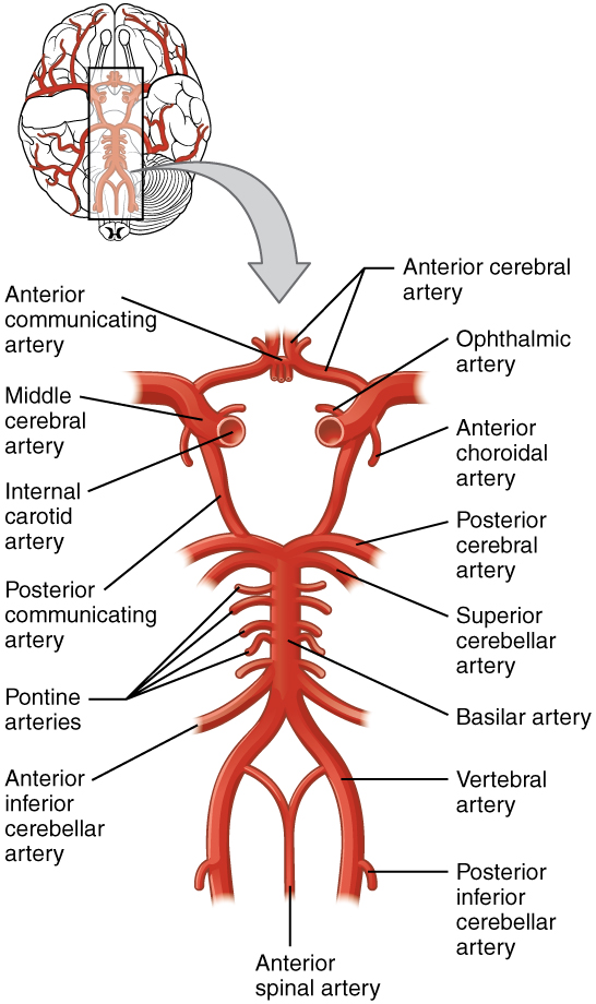
The major artery carrying recently oxygenated blood away from the heart is the aorta. The very first branches off the aorta supply the heart with nutrients and oxygen. The next branches give rise to the common carotid arteries, which further branch into the internal carotid arteries. The external carotid arteries supply blood to the tissues on the surface of the cranium. A second set of vessels that supply the CNS are the vertebral arteries. Branches off the left and right vertebral arteries merge into the anterior spinal artery supplying the anterior aspect of the spinal cord. The two vertebral arteries then merge into the basilar artery. The left and right internal carotid arteries and branches of the basilar artery all become the circle of Willis, an anastomosis or confluence of arteries that can maintain perfusion of the brain even if narrowing or a blockage limits flow through one part (Figure 6.2.1).
External Website

Watch AnatomyZone’s animation [YouTube] to see how blood flows to the brain and passes through the circle of Willis before being distributed through the cerebrum. The circle of Willis is a specialized arrangement of arteries that ensure constant perfusion of the cerebrum even in the event of a blockage of one of the arteries in the circle. The animation shows the normal direction of flow through the circle of Willis to the middle cerebral artery.
Venous Return
After passing through the CNS, blood returns to the circulation through a series of dural sinuses (channels) and veins (Figure 6.2.2). The superior sagittal sinus runs in the groove of the longitudinal fissure, where it absorbs CSF from the meninges. The superior sagittal sinus drains to the confluence of sinuses to then drain into the transverse sinuses. The transverse sinuses connect to the sigmoid sinuses, which then connect to the jugular veins. From there, the blood continues toward the heart to be pumped to the lungs for reoxygenation.

Cerebrovascular Accidents
Damage to the nervous system can be limited to individual structures or can be distributed across broad areas of the brain and spinal cord. Localized, limited injury to the nervous system is most often the result of circulatory problems. Neurons are very sensitive to oxygen deprivation and will start to deteriorate within one or two minutes, and permanent damage (cell death) could result within a few hours. The loss of blood flow to part of the brain is known as a stroke, or a cerebrovascular accident (CVA).
There are two main types of stroke, depending on how the blood supply is compromised: ischemic and hemorrhagic. An ischemic stroke is the loss of blood flow to an area because vessels are blocked or narrowed. This is often caused by an embolus, which may be a blood clot or fat deposit. Ischemia may also be the result of thickening of the blood vessel wall or a drop in blood volume in the brain known as hypovolemia.
A related type of CVA is known as a transient ischemic attack (TIA), which is similar to a stroke, although it does not last as long. The diagnostic definition of a stroke includes effects that last at least 24 hours. Any stroke symptoms that are resolved within a 24-hour period because of restoration of adequate blood flow are classified as a TIA.
A hemorrhagic stroke is bleeding into the brain because of a damaged blood vessel. Accumulated blood fills a region of the cranial vault and presses against the tissue in the brain (Figure 6.2.3). Physical pressure on the brain can cause the loss of function, as well as the squeezing of local arteries resulting in compromised blood flow beyond the site of the hemorrhage. As blood pools in the nervous tissue and the vasculature is damaged, the blood-brain barrier can break down and allow additional fluid to accumulate in the region, which is known as edema.
Protective Coverings of the Brain and Spinal Cord
The skull is a protective bone cavity which covers the brain. In addition, the outer surface of the CNS is covered by a series of membranes composed of connective tissue called the meninges, which protect the brain. The dura mater is a thick fibrous layer and a strong protective sheath over the entire brain and spinal cord. It is anchored to the inner surface of the cranium and vertebral cavity. The arachnoid mater is a membrane of thin fibrous tissue that forms a loose sac around the CNS. Directly adjacent to the surface of the CNS is the pia mater, a thin fibrous membrane that follows the convolutions of gyri and sulci in the cerebral cortex and fits into other grooves and indentations (Figure 6.2.4).
Dura Mater
Like a thick cap covering the brain, the dura mater is a tough outer covering. The name comes from the Latin for “tough mother” to represent its physically protective role. It encloses the entire CNS and the major blood vessels that enter the cranium and vertebral cavity. It is directly attached to the inner surface of the bones of the cranium and to the very end of the vertebral cavity.
Arachnoid Mater
The middle layer of the meninges is the arachnoid, named for the spiderweb-like trabeculae between it and the pia mater. The arachnoid defines a sac-like enclosure around the CNS. The subarachnoid space is filled with circulating CSF. The arachnoid emerges into the dural sinuses as the arachnoid granulations, where the CSF is filtered back into the blood for drainage from the nervous system.
The subarachnoid space is filled with circulating CSF, which also provides a liquid cushion to the brain and spinal cord. Similar to clinical blood work, a sample of CSF can be withdrawn to find chemical evidence of neuropathology or metabolic traces of the biochemical functions of nervous tissue.
Pia Mater
The outer surface of the CNS is covered in the thin fibrous membrane of the pia mater. It is thought to have a continuous layer of cells providing a fluid-impermeable membrane. The name pia mater comes from the Latin for “tender mother,” suggesting the thin membrane is a gentle covering for the brain. The pia extends into every convolution of the CNS, lining the inside of the sulci in the cerebral and cerebellar cortices. At the end of the spinal cord, a thin filament extends from the inferior end of CNS at the upper lumbar region of the vertebral column to the sacral end of the vertebral column. Because the spinal cord does not extend through the lower lumbar region of the vertebral column, a needle can be inserted through the dura and arachnoid layers to withdraw CSF. This procedure is called a lumbar puncture and avoids the risk of damaging the central tissue of the spinal cord. Blood vessels that are nourishing the central nervous tissue are between the pia mater and the nervous tissue.
Disorders of the Meninges
Meningitis is an inflammation of the meninges, the three layers of fibrous membrane that surround the CNS. Meningitis can be caused by infection by bacteria or viruses. The particular pathogens are not special to meningitis; it is just an inflammation of that specific set of tissues from what might be a broader infection. Bacterial meningitis can be caused by Streptococcus, Staphylococcus, or the tuberculosis pathogen, among many others. Viral meningitis is usually the result of common enteroviruses (such as those that cause intestinal disorders) but may be the result of the herpes virus or West Nile virus. Bacterial meningitis tends to be more severe.
The symptoms associated with meningitis can be fever, chills, nausea, vomiting, light sensitivity, soreness of the neck, or severe headache. More important are the neurological symptoms, such as changes in mental state (confusion, memory deficits, and other dementia-type symptoms). A serious risk of meningitis can be damage to peripheral structures because of the nerves that pass through the meninges. Hearing loss is a common result of meningitis.
The primary test for meningitis is a lumbar puncture. A needle inserted into the lumbar region of the spinal column through the dura mater and arachnoid membrane into the subarachnoid space can be used to withdraw the fluid for chemical testing. Fatality occurs in 5% to 40% of children and 20% to 50% of adults with bacterial meningitis. Treatment of bacterial meningitis is through antibiotics, but viral meningitis cannot be treated with antibiotics because viruses do not respond to that type of drug. Fortunately, the viral forms are milder.
External Website

Watch this Medical Centric’s video [YouTube} that describes the procedure known as the lumbar puncture, a medical procedure used to sample the CSF. Because of the anatomy of the CNS, it is a relative safe location to insert a needle.
The Ventricular System
Cerebrospinal fluid (CSF) circulates throughout and around the CNS. In other tissues, water and small molecules are filtered through capillaries as the major contributor to the interstitial fluid. In the brain, CSF is produced in special structures to perfuse through the nervous tissue of the CNS and is continuous with the interstitial fluid. Specifically, CSF circulates to remove metabolic wastes from the interstitial fluids of nervous tissues and return them to the blood stream. The ventricles are the open spaces within the brain where CSF circulates. In some of these spaces, CSF is produced by filtering of the blood that is performed by a specialized membrane known as a choroid plexus. The CSF circulates through all of the ventricles to eventually emerge into the subarachnoid space where it will be reabsorbed into the blood.
The Ventricles
There are four ventricles within the brain, all of which developed from the original hollow space within the neural tube, the central canal. The first two are named the lateral ventricles and are deep within the cerebrum. These ventricles are connected to the third ventricle by two openings. The third ventricle is the space between the left and right sides of the diencephalon, which opens into the cerebral aqueduct that passes through the midbrain. The aqueduct opens into the fourth ventricle, which is the space between the cerebellum and the pons and upper medulla (Figure 6.2.5).
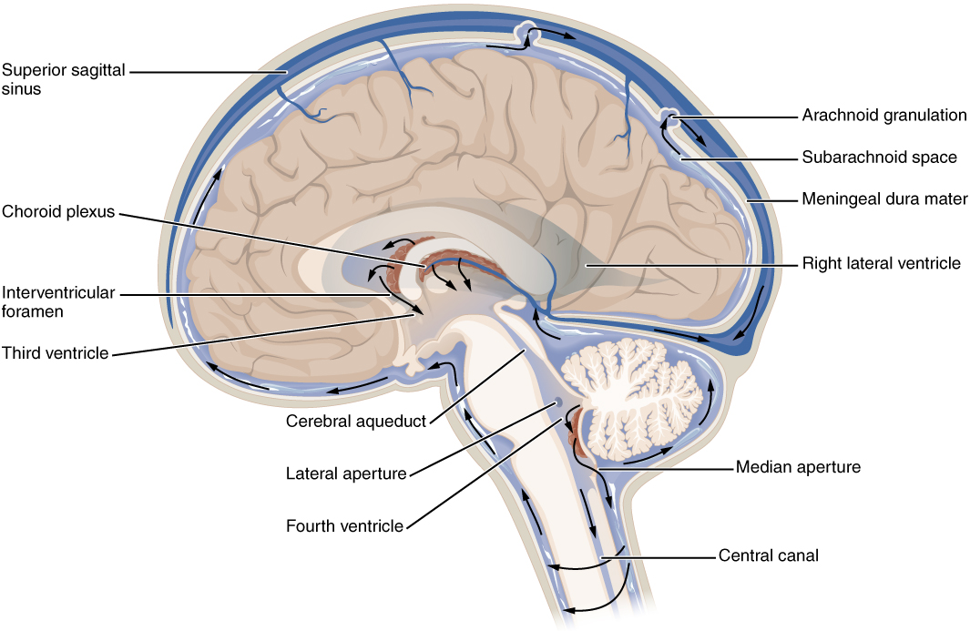
The ventricular system opens up to the subarachnoid space from the fourth ventricle. The single median aperture and the pair of lateral apertures connect to the subarachnoid space so that CSF can flow through the ventricles and around the outside of the CNS. Cerebrospinal fluid is produced within the ventricles by a type of specialized membrane called a choroid plexus. Ependymal cells (one of the types of glial cells described in the introduction to the nervous system) surround blood capillaries and filter the blood to make CSF. The fluid is a clear solution with a limited amount of the constituents of blood. It is essentially water, small molecules, and electrolytes. Oxygen and carbon dioxide are dissolved into the CSF, as they are in blood, and can diffuse between the fluid and the nervous tissue.
Cerebrospinal Fluid Circulation
The choroid plexuses are found in all four ventricles. Observed in dissection, they appear as soft, fuzzy structures that may still be pink, depending on how well the circulatory system is cleared in preparation of the tissue. The CSF is produced from components extracted from the blood, so its flow out of the ventricles is tied to the pulse of cardiovascular circulation.
From the lateral ventricles, the CSF flows into the third ventricle, where more CSF is produced, and then through the cerebral aqueduct into the fourth ventricle where even more CSF is produced. A very small amount of CSF is filtered at any one of the plexuses, for a total of about 500 milliliters daily, but it is continuously made and pulses through the ventricular system, keeping the fluid moving. From the fourth ventricle, CSF can continue down the central canal of the spinal cord, but this is essentially a cul-de-sac, so more of the fluid leaves the ventricular system and moves into the subarachnoid space through the median and lateral apertures.
Within the subarachnoid space, the CSF flows around all of the CNS, providing two important functions. As with elsewhere in its circulation, the CSF picks up metabolic wastes from the nervous tissue and moves it out of the CNS. It also acts as a liquid cushion for the brain and spinal cord. By surrounding the entire system in the subarachnoid space, it provides a thin buffer around the organs within the strong, protective dura mater. The arachnoid granulations are outpocketings of the arachnoid membrane into the dural sinuses so that CSF can be reabsorbed into the blood, along with the metabolic wastes. From the dural sinuses, blood drains out of the head and neck through the jugular veins, along with the rest of the circulation for blood, to be reoxygenated by the lungs and wastes to be filtered out by the kidneys (Table 6.2).
| Description | Lateral Ventricles | Third Ventricle | Cerebral Aqueduct | Fourth Ventricle | Central Canal | Subarachnoid Space |
|---|---|---|---|---|---|---|
| Location in CNS | Cerebrum | Diencephalon | Midbrain | Between pons/upper medulla and cerebellum | Spinal cord | External to entire CNS |
| Blood vessel structure | Choroid plexus | Choroid plexus | None | Choroid plexus | None | Arachnoid granulations |
Section Review
The CNS has a privileged blood supply established by the blood-brain barrier. Establishing this barrier are anatomical structures that help to protect and isolate the CNS. The arterial blood to the brain comes from the internal carotid and vertebral arteries, which both contribute to the unique circle of Willis that provides constant perfusion of the brain even if one of the blood vessels is blocked or narrowed. That blood is eventually filtered to make a separate medium, the CSF, that circulates within the spaces of the brain and then into the surrounding space defined by the meninges, the protective covering of the brain and spinal cord.
The blood that nourishes the brain and spinal cord is behind the glial cell–enforced blood-brain barrier, which limits the exchange of material from blood vessels with the interstitial fluid of the nervous tissue. Thus, metabolic wastes are collected in cerebrospinal fluid that circulates through the CNS. This fluid is produced by filtering blood at the choroid plexuses in the four ventricles of the brain. It then circulates through the ventricles and into the subarachnoid space, between the pia mater and the arachnoid mater. From the arachnoid granulations, CSF is reabsorbed into the blood, removing the waste from the privileged central nervous tissue.
The blood, now with the reabsorbed CSF, drains out of the cranium through the dural sinuses. The dura mater is the tough outer covering of the CNS, which is anchored to the inner surface of the cranial and vertebral cavities. It surrounds the venous space known as the dural sinuses, which connect to the jugular veins, where blood drains from the head and neck.
Review Questions
Critical Thinking Questions
Glossary
- anastomosis
- area where vessels unite to form interconnections that normally allow blood to circulate to a region even if there may be partial blockage in another branch.
- anterior spinal artery
- blood vessel from the merged branches of the vertebral arteries that runs along the anterior surface of the spinal cord
- arachnoid granulation
- outpocket of the arachnoid membrane into the dural sinuses that allows for reabsorption of CSF into the blood
- arachnoid mater
- middle layer of the meninges named for the spiderweb-like trabeculae that extend between it and the pia mater
- basilar artery
- blood vessel from the merged vertebral arteries that runs along the dorsal surface of the brain stem
- central canal
- hollow space within the spinal cord that is the remnant of the center of the neural tube
- cerebral aqueduct
- connection of the ventricular system between the third and fourth ventricles located in the midbrain
- choroid plexus
- specialized structures containing ependymal cells lining blood capillaries that filter blood to produce CSF in the four ventricles of the brain
- circle of Willis
- unique anatomical arrangement of blood vessels around the base of the brain that maintains perfusion of blood into the brain even if one component of the structure is blocked or narrowed
- common carotid artery
- blood vessel that branches off the aorta (or the brachiocephalic artery on the right) and supplies blood to the head and neck
- dura mater
- tough, fibrous outer layer of the meninges that is attached to the inner surface of the cranium and vertebral column and surrounds the entire CNS
- dural sinus
- any of the venous structures surrounding the brain, enclosed within the dura mater, which drain blood from the CNS to the common venous return of the jugular veins
- fourth ventricle
- the portion of the ventricular system that is in the region of the brain stem and opens into the subarachnoid space through the median and lateral apertures
- internal carotid artery
- branch from the common carotid artery that enters the cranium and supplies blood to the brain
- jugular veins
- blood vessels that return “used” blood from the head and neck
- lateral apertures
- pair of openings from the fourth ventricle to the subarachnoid space on either side and between the medulla and cerebellum
- lateral ventricles
- portions of the ventricular system that are in the region of the cerebrum
- lumbar puncture
- procedure used to withdraw CSF from the lower lumbar region of the vertebral column that avoids the risk of damaging CNS tissue because the spinal cord ends at the upper lumbar vertebrae
- median aperture
- singular opening from the fourth ventricle into the subarachnoid space at the midline between the medulla and cerebellum
- meninges
- protective outer coverings of the CNS composed of connective tissue
- pia mater
- thin, innermost membrane of the meninges that directly covers the surface of the CNS
- sigmoid sinuses
- dural sinuses that drain directly into the jugular veins
- subarachnoid space
- space between the arachnoid mater and pia mater that contains CSF and the fibrous connections of the arachnoid trabeculae
- superior sagittal sinus
- dural sinus that runs along the top of the longitudinal fissure and drains blood from the majority of the outer cerebrum
- third ventricle
- portion of the ventricular system that is in the region of the diencephalon
- transverse sinuses
- dural sinuses that drain along either side of the occipital–cerebellar space
- ventricles
- remnants of the hollow center of the neural tube that are spaces for cerebrospinal fluid to circulate through the brain
- vertebral arteries
- arteries that ascend along either side of the vertebral column through the transverse foramina of the cervical vertebrae and enter the cranium through the foramen magnum
Glossary Flashcards
This work, Human Physiology, is adapted from Anatomy & Physiology by OpenStax, licensed under CC BY. This edition, with revised content and artwork, is licensed under CC BY-SA except where otherwise noted.
Images from Anatomy & Physiology by OpenStax are licensed under CC BY except where otherwise noted.
Access the original for free at OpenStax.
Image Descriptions
Figure 6.2.1. This anatomical diagram shows the arterial system that supplies blood to the brain, viewed from underneath. The illustration depicts how blood flows upward through paired vertebral arteries that merge into a single basilar artery along the brainstem. The basilar artery branches into posterior cerebral arteries. Separately, paired internal carotid arteries rise on either side and branch into middle cerebral arteries and anterior cerebral arteries. These major vessels are interconnected by smaller communicating arteries, forming a roughly circular structure called the Circle of Willis at the base of the brain. Additional smaller branches extend to supply the cerebellum, brainstem, eyes, and spinal cord. This interconnected network ensures the brain receives continuous blood flow even if one vessel becomes blocked. Red coloring indicates oxygen-rich arterial blood throughout the system. [Return to Figure 6.2.1]
Figure 6.2.2. This anatomical diagram shows a side view cross-section of the human head, revealing the venous sinuses that drain blood from the brain. The illustration depicts layers from outside to inside: the cranium (skull bone) shown in peach, the dura mater (a tough protective membrane) in gray beneath it, and the brain tissue in beige filling the center. Between the dura mater layers run large venous channels called sinuses, shown in blue. Along the top of the brain runs the superior sagittal sinus, which curves down the back to meet the straight sinus running horizontally through the middle. These converge at the confluence of sinuses at the back of the head. Below this junction, the transverse sinus runs along the back and side, continuing as the sigmoid sinus (S-shaped) which eventually drains into the jugular vein in the neck. The inferior sagittal sinus runs along the lower inner surface, and the occipital sinus is located at the back. Smaller cerebral veins feed into the great cerebral vein, which connects to this sinus system. The lower portion shows the nasal cavity, teeth, and neck musculature in coral-red. Blue arrows indicate the direction of blood flow through these venous channels. [Return to Figure 6.2.2]
Figure 6.2.3. This image presents two side-by-side views of the same brain cross-section, both looking down from above. Image (a) on the left is a simplified anatomical illustration showing the brain’s outline with skull represented by black lines. The brain tissue appears in beige tones, with a large dark red region occupying the left side of the brain, representing a hemorrhage or bleed. This abnormal area has irregular, cloud-like borders and extends roughly one-quarter to one-third across the brain from left to right. Image (b) on the right shows an actual CT scan of the same condition. In this grayscale medical image, the skull appears as a bright white ring, normal brain tissue shows as gray, and the hemorrhage appears as a bright white irregular mass in the corresponding left-side location. The CT scan also reveals some darker areas within and around the bright hemorrhage, indicating blood of different densities or ages. Both images demonstrate how bleeding in the brain displaces normal tissue and creates a mass effect, with the illustrated version simplifying the pathology for educational purposes while the CT scan shows the actual clinical appearance. [Return to Figure 6.2.3]
Figure 6.2.4. This anatomical diagram shows a detailed cross-section of the layers covering and protecting the brain, viewed from the side. From outermost to innermost, the structures include: the bone (skull) at the top, shown in beige with a porous, spongy texture; the dura mater, a thick tough membrane shown in blue-gray directly beneath the skull; the subdural space, a thin potential space between membranes; the arachnoid mater, a delicate membrane shown in orange; the subarachnoid space, a fluid-filled gap containing cerebrospinal fluid; and the pia mater, a thin membrane shown in orange that adheres directly to the brain surface. Within the subarachnoid space are visible arachnoid trabeculae, which are delicate connective tissue strands bridging between the arachnoid and pia mater. The diagram also shows arachnoid granulation villi, which are finger-like projections extending upward from the arachnoid layer into the superior sagittal sinus (a large venous channel within the dura mater shown in blue at the top center). These villi allow cerebrospinal fluid to drain back into the bloodstream. Blue veins are visible coursing through and along these layers. At the bottom, the cerebral cortex (brain tissue) is shown in beige with its characteristic folded surface, and the longitudinal fissure (the deep groove separating the brain’s two hemispheres) is visible as a vertical cleft in the center. Orange-colored cerebral veins can be seen within the brain tissue [Return to Figure 6.2.4]
Figure 6.2.5. This diagram presents a side view cross-section of the brain, illustrating the cerebrospinal fluid (CSF) circulation system. The brain tissue appears in beige with its characteristic folded surface and internal structures visible. Surrounding the brain are the protective membranes: the meningeal dura mater (shown in tan-beige) lines the inside of the skull, followed by the subarachnoid space (shown in light blue) filled with cerebrospinal fluid, which cushions the entire brain surface. Arachnoid granulations, small bulb-like projections where CSF drains into the bloodstream, protrude into the superior sagittal sinus, a large blue venous channel running along the top of the brain. The diagram highlights the brain’s internal fluid-filled chambers called ventricles, shown in blue. These include the right lateral ventricle, a curved C-shaped cavity within the cerebral hemisphere; the third ventricle, a narrow midline chamber in the center of the brain; and the fourth ventricle, located at the back lower portion of the brain, which has a distinctive tree-like structure (the cerebellum) shown in white behind it. These ventricles connect through narrow passages: the interventricular foramen (also called foramen of Monro) connects the lateral ventricle to the third ventricle, and the cerebral aqueduct, a thin channel, connects the third to the fourth ventricle. Black arrows throughout the diagram indicate CSF flow direction—from the choroid plexus (red vascular structures inside the ventricles where CSF is produced), through the ventricular system, and out through openings called the lateral aperture and median aperture at the fourth ventricle into the subarachnoid space. The central canal, a thin fluid channel extending downward into the spinal cord, is also labeled at the bottom of the diagram. [Return to Figure 6.2.5]
Report an Error
Did you find an error, typo, broken link, or other problem in the text? Please follow this link to the error reporting form to submit an error report to the authors.
