Chapter 6. The Central Nervous System
6.4 The Cerebrum
Learning Objectives
By the end of this section, you will be able to:
- List the four major regions of the adult brain
- List the basic regions of the cerebrum
- List the major lobes of the cerebral cortex and describe their general function
- Recall the different types of memory
- Compare and contrast Broca’s and Wernicke’s aphasia in terms of their symptoms
- Recall the brain region associated with judgment and abstract reasoning
- Describe the functions of the amygdala, hippocampus, and basal nuclei
The brain and the spinal cord are the central nervous system, and they represent the main organs of the nervous system. The spinal cord is a single structure, whereas the adult brain is described in terms of four major regions: the cerebrum, the diencephalon, the brain stem, and the cerebellum. A person’s conscious experiences are based on neural activity in the brain. The regulation of homeostasis is governed by a specialized region in the brain. The coordination of reflexes depends on the integration of sensory and motor pathways in the spinal cord.

The Cerebrum
The iconic gray mantle of the human brain, which appears to make up most of the mass of the brain, is the cerebrum (Figure 6.4.2). The cerebrum consists of the outer layer known as the cerebral cortex and subcortical structures beneath the outer layer. There is a large separation between the two sides of the cerebrum called the longitudinal fissure. It separates the cerebrum into two distinct halves: a right and left cerebral hemisphere. Deep within the cerebrum, the white matter of the corpus callosum provides the major pathway for communication between the two hemispheres of the cerebral cortex.

Many of the higher neurological functions, such as memory, emotion, and consciousness, are the result of cerebral function. The complexity of the cerebrum is different across vertebrate species. The cerebrum of the most primitive vertebrates is not much more than the connection for the sense of smell. In mammals, the cerebrum comprises the outer gray matter that is the cortex (from the Latin word meaning “bark of a tree”) and several deep nuclei that belong to three important functional groups. The basal nuclei are responsible for cognitive processing, the most important function being that associated with planning movements. The basal forebrain contains nuclei that are important in learning and memory. The limbic cortex is the region of the cerebral cortex that is part of the limbic system, a collection of structures involved in emotion, memory, and behavior.
Cerebral Cortex
The cerebrum is covered by a continuous layer of gray matter that wraps around either side of the forebrain—the cerebral cortex. This thin, extensive region of wrinkled gray matter is responsible for the higher functions of the nervous system. The pattern of these folds of tissue indicates specific regions of the cerebral cortex. The head is limited by the size of the birth canal, and the brain must fit inside the cranial cavity of the skull. Extensive folding in the cerebral cortex enables more gray matter to fit into this limited space. If the gray matter of the cortex were peeled off of the cerebrum and laid out flat, its surface area would be roughly equal to one square meter.
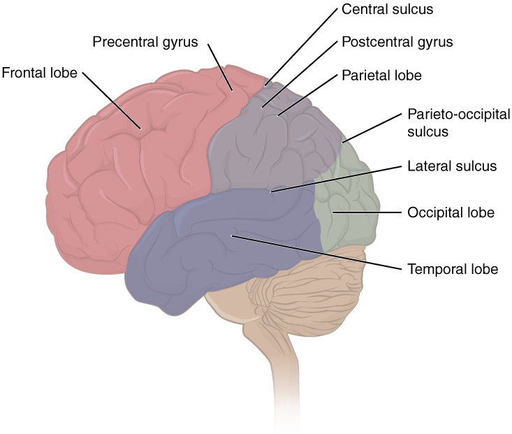
The surface of the brain can be mapped on the basis of the locations of large gyri and sulci. Using these landmarks, the cortex can be separated into four major regions, or lobes (Figure 6.4.3). The lateral sulcus that separates the temporal lobe from the other regions is one such landmark. Superior to the lateral sulcus are the parietal lobe and frontal lobe, which are separated from each other by the central sulcus. The posterior region of the cortex is the occipital lobe, which has no obvious anatomical border between it and the parietal or temporal lobes on the lateral surface of the brain. From the medial surface, an obvious landmark separating the parietal and occipital lobes is called the parieto-occipital sulcus. The fact that there is no obvious anatomical border between these lobes is consistent with the functions of these regions being interrelated.
Anterior to the central sulcus is the frontal lobe, which is primarily associated with motor functions and planning. The frontal lobe includes the primary motor cortex, the premotor area/cortex, the frontal eye fields, Broca’s area, the prefrontal cortex, and other regions as well. Cells within the primary motor cortex are the upper motor neurons that instruct cells in the spinal cord and brain stem (lower motor neurons) to move skeletal muscles. The premotor area is responsible for storing learned movement algorithms, which are instructions for complex movements. Different algorithms activate the upper motor neurons in the correct sequence when a complex motor activity is performed. The frontal eye fields are important in eliciting scanning eye movements and in attending to visual stimuli. Broca’s area is responsible for the production of language, or controlling movements responsible for speech; in the vast majority of people, it is located only on the left side. The prefrontal cortex serves cognitive functions that can be the basis of personality, short-term memory, and consciousness. The prefrontal lobotomy is an outdated mode of treatment for personality disorders (psychiatric conditions) that profoundly affected the personality of the patient.
The main sensation associated with the parietal lobe is somatosensation, meaning the general sensations associated with the body. Within the parietal lobe is the primary somatosensory cortex. All of the tactile senses are processed in this area, including touch, pressure, tickle, pain, itch, and vibration, as well as more general senses of the body such as proprioception and kinesthesia, which are the senses of body position and movement, respectively.
The occipital lobe is responsible for primary visual perception. That visual information is complex, so it is processed in the temporal and parietal lobes as well.
The temporal lobe is associated with primary auditory sensation. Because regions of the temporal lobe are part of the limbic system, memory is an important function associated with that lobe. Structures in the temporal lobe are responsible for establishing long-term memory, but the ultimate location of those memories is usually in the region in which the sensory perception was processed.
Functions of the Cerebral Cortex
The cerebrum is the seat of many of the higher mental functions, such as memory and learning, language, and conscious perception, which are the subjects of subtests of the mental status exam. The cerebral cortex is the thin layer of gray matter on the outside of the cerebrum. It is approximately a millimeter thick in most regions and highly folded to fit within the limited space of the cranial vault. These higher functions are distributed across various regions of the cortex, and specific locations can be said to be responsible for particular functions. There is a limited set of regions, for example, that are involved in language function, and they can be subdivided on the basis of the particular part of language function that each governs.

The primary cortical areas are where sensory information is initially processed, or where motor commands emerge to go to the brain stem or spinal cord. Association areas are adjacent to primary areas and further process the modality-specific input. A number of other regions, which extend beyond these primary or association areas of the cortex, are referred to as integrative areas. These areas are found in the spaces between the domains for particular sensory or motor functions, and they integrate multisensory information, or process sensory or motor information in more complex ways. Consider, for example, the posterior parietal cortex that lies between the somatosensory cortex and visual cortex regions. This has been ascribed to the coordination of visual and motor functions, such as reaching to pick up a glass. The somatosensory function that would be part of this is the proprioceptive feedback from moving the arm and hand. The weight of the glass, based on what it contains, will influence how those movements are executed.
Cognitive Abilities
Assessment of cerebral functions is directed at cognitive abilities. The abilities can be separated into four groups: orientation and memory, language and speech, sensorium, and judgment and abstract reasoning.
Orientation and Memory
Orientation is the patient’s awareness of their immediate circumstances. It is awareness of time, not in terms of the clock, but of the date and what is occurring around the patient. It is awareness of place, such that a patient should know where he or she is and why. It is also awareness of who the patient is—recognizing personal identity and being able to relate that to the examiner.
Memory is largely a function of the temporal lobe, along with structures beneath the cerebral cortex such as the hippocampus and the amygdala. The storage of memory requires these structures of the medial temporal lobe. A famous case of a man who had both medial temporal lobes removed to treat intractable epilepsy provided insight into the relationship between the structures of the brain and the function of memory.
Henry Molaison, who was referred to as patient HM when he was alive, had epilepsy localized to both of his medial temporal lobes. In 1953, a bilateral lobectomy was performed that alleviated the epilepsy but resulted in the inability for HM to form new memories—a condition called anterograde amnesia. HM was able to recall most events from before his surgery, although there was a partial loss of earlier memories, which is referred to as retrograde amnesia. HM became the subject of extensive studies into how memory works. What he was unable to do was form new memories of what happened to him, what are now called episodic memory. Episodic memory is autobiographical in nature, such as remembering riding a bicycle as a child around the neighborhood, as opposed to the procedural memory of how to ride a bike. HM also retained his short-term memory, also known as working memory, which temporarily holds information for a few seconds. After a brief period, those memories would dissipate or decay and not be stored long term because the medial temporal lobe structures were removed.
The difference in short-term, procedural, and episodic memory, as evidenced by patient HM, suggests that there are different parts of the brain responsible for those functions. The long-term storage of episodic memory requires the hippocampus and related medial temporal structures, and the location of those memories is in the multimodal integration areas of the cerebral cortex. However, short-term memory—also called working or active memory—is localized to the prefrontal lobe. Because patient HM had only lost his medial temporal lobe—and lost very little of his previous memories and did not lose the ability to form new short-term memories—it was concluded that the function of the hippocampus, and adjacent structures in the medial temporal lobe, is to move (or consolidate) short-term memories (in the prefrontal lobe) to long-term memory (in the temporal lobe).
Language and Speech
Language is, arguably, a very human aspect of neurological function. The neurological exam has two specific subtests that address language. One measures the ability of the patient to understand language by asking them to follow a set of instructions to perform an action, such as “touch your right finger to your left elbow and then to your right knee.” Another subtest assesses the fluency and coherency of language by having the patient generate descriptions of objects or scenes depicted in drawings and recite sentences or explain a written passage. Language, however, is important in so many ways in the neurological exam. The patient needs to know what to do, whether it is as simple as explaining how the knee-jerk reflex is going to be performed or asking a question such as “What is your name?” Often, language deficits can be determined without specific subtests; if a person cannot reply to a question properly, there may be a problem with the reception of language.
An important example of multimodal integrative areas is associated with language function (Figure 6.4.5). Adjacent to the auditory association cortex, at the end of the lateral sulcus just anterior to the visual cortex, is Wernicke’s area. In the lateral aspect of the frontal lobe, just anterior to the region of the motor cortex associated with the head and neck, is Broca’s area. Both regions were originally described on the basis of losses of speech and language, which is called aphasia. The aphasia associated with Broca’s area is known as an expressive aphasia, which means that speech production is compromised. This type of aphasia is often described as non-fluency because the ability to say some words leads to broken or halting speech. Grammar can also appear to be lost. The aphasia associated with Wernicke’s area is known as a receptive aphasia, which is not a loss of speech production but a loss of understanding of content. Patients, after recovering from acute forms of this aphasia, report not being able to understand what is said to them or what they are saying themselves, but they often cannot keep from talking.
The two regions are connected by white matter tracts that run between the posterior temporal lobe and the lateral aspect of the frontal lobe. Conduction aphasia associated with damage to this connection refers to the problem of connecting the understanding of language to the production of speech. This is a very rare condition, but it is likely to present as an inability to faithfully repeat spoken language.
External Website


Sensorium
Those parts of the brain involved in the reception and interpretation of sensory stimuli are referred to collectively as the sensorium. The cerebral cortex has several regions that are necessary for sensory perception. From the primary cortical areas of the somatosensory, visual, auditory, and gustatory senses to the association areas that process information in these modalities, the cerebral cortex is the seat of conscious sensory perception. In contrast, sensory information can also be processed by deeper brain regions, which we may vaguely describe as subconscious—for instance, we are not constantly aware of the proprioceptive information that the cerebellum uses to maintain balance.
Judgment and Abstract Reasoning
Planning and producing responses requires an ability to make sense of the world around us. Making judgments and reasoning in the abstract are necessary to produce movements as part of larger responses. For example, when your alarm goes off, do you hit the snooze button or jump out of bed? Is 10 extra minutes in bed worth the extra rush to get ready for your day? Will hitting the snooze button multiple times lead to feeling more rested or result in a panic as you run late? How you mentally process these questions can affect your whole day.
The prefrontal cortex is responsible for the functions responsible for planning and making decisions. The prefrontal cortex is composed of the regions of the frontal lobe that are not directly related to specific motor functions. The most posterior region of the frontal lobe, the precentral gyrus, is the primary motor cortex. Anterior to that are the premotor cortex, Broca’s area, and the frontal eye fields, which are all related to planning certain types of movements. Anterior to what could be described as motor association areas are the regions of the prefrontal cortex. They are the regions in which judgment, abstract reasoning, and working memory are localized. The antecedents to planning certain movements are judging whether those movements should be made, as in the example of deciding whether to hit the snooze button.
To an extent, the prefrontal cortex may be related to personality. The neurological exam does not necessarily assess personality, but it can be within the realm of neurology or psychiatry. A clinical situation that suggests this link between the prefrontal cortex and personality comes from the story of Phineas Gage, the railroad worker from the mid-1800s who had a metal spike impale his prefrontal cortex. There are suggestions that the steel rod led to changes in his personality. A man who was a quiet, dependable railroad worker became a raucous, irritable drunkard. Later anecdotal evidence from his life suggests that he was able to support himself, although he had to relocate and take on a different career as a stagecoach driver.
A psychiatric practice to deal with various disorders was the prefrontal lobotomy. This procedure was common in the 1940s and early 1950s until antipsychotic drugs became available. The connections between the prefrontal cortex and other regions of the brain were severed. The disorders associated with this procedure included some aspects of what are now referred to as personality disorders but also included mood disorders and psychoses. Depictions of lobotomies in popular media suggest a link between cutting the white matter of the prefrontal cortex and changes in a patient’s mood and personality, though this correlation is not well understood.
Everyday Connections – The Myth of Left Brain/Right Brain
There is a persistent myth that people are “right-brained” or “left-brained,” which is an oversimplification of an important concept about the cerebral hemispheres. There is some lateralization of function, in which the left side of the brain is devoted to language function and the right side is devoted to spatial and nonverbal reasoning. Whereas these functions are predominantly associated with those sides of the brain, there is no monopoly by either side on these functions. Many pervasive functions, such as language, are distributed globally around the cerebrum.
Some of the support for this misconception has come from studies of split brains. A drastic way to deal with a rare and devastating neurological condition (intractable epilepsy) is to separate the two hemispheres of the brain. After sectioning the corpus callosum, a split-brained patient will have trouble producing verbal responses on the basis of sensory information processed on the right side of the cerebrum, leading to the idea that the left side is responsible for language function.
However, there are well-documented cases of language functions lost from damage to the right side of the brain. The deficits seen in damage to the left side of the brain are classified as aphasia, a loss of speech function; damage on the right side can affect the use of language. Right-side damage can result in a loss of ability to understand figurative aspects of speech, such as jokes, irony, or metaphors. Nonverbal aspects of speech can be affected by damage to the right side, such as facial expression or body language, and right-side damage can lead to a “flat affect” in speech, or a loss of emotional expression in speech—in other words, sounding like a robot when talking. Damage to language areas on the right side causes a condition called aprosodia where the patient has difficulty understanding or expressing the figurative parts of speech.
Subcortical Structures
Beneath the cerebral cortex are sets of nuclei known as subcortical nuclei that augment cortical processes. The nuclei of the basal forebrain serve as the primary location for acetylcholine production, which modulates the overall activity of the cortex, possibly leading to greater attention to sensory stimuli. Alzheimer’s disease is associated with a loss of neurons in the basal forebrain. The hippocampus and amygdala are medial-lobe structures that, along with the adjacent cortex, are involved in long-term memory formation and emotional responses. The basal nuclei are a set of nuclei in the cerebrum responsible for comparing cortical processing with the general state of activity in the nervous system to influence the likelihood of movement taking place. For example, while a student is sitting in a classroom listening to a lecture, the basal nuclei will keep the urge to jump up and scream from actually happening. (The basal nuclei are also referred to as the basal ganglia, although that is potentially confusing because the term ganglia is typically used for peripheral structures.)
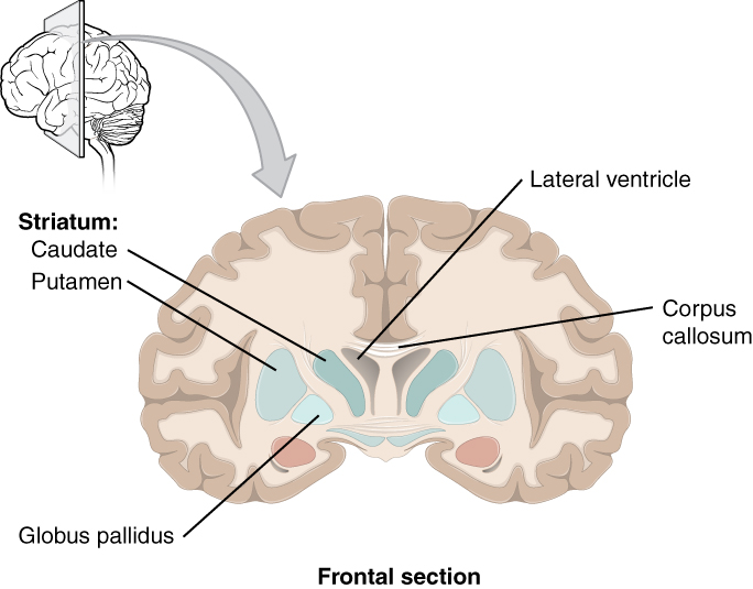
The major structures of the basal nuclei that control movement are the caudate, putamen, and globus pallidus, which are located deep in the cerebrum. Together, the caudate and putamen are called the striatum. The globus pallidus has two subdivisions, the external and internal segments, which are lateral and medial, respectively. These nuclei are depicted in a frontal section of the brain in Figure 6.4.6.
Measuring Brain Activity
The electroencephalogram (EEG) is one method by which we can observe electrical activity of the brain. When neurons send action potentials, several charged ions move across cell membranes, which causes a change in the electrical charge of the adjacent area. Some of these charges are very large, especially when several millions of neurons are sending action potentials at the same time. These currents produced by neurons of the outer surface of the brain (cortex) can be so large that they are detectable outside the skull at the surface of the head. The EEG works by recording these changes at different areas of the brain.
The theory behind the EEG is the same used in an electrocardiogram (ECG) to detect the electrical activity of heart cells. To perform an EEG, a gel is applied onto small patches of the scalp. This gel is basically a sodium chloride gel that acts as a conductor, allowing the currents to be picked up by a series of adhesive electrodes. Each electrode is placed on a specific area of the scalp, and the information from each electrode runs into a computer. From there, the computer can then perform calculations to determine which cortical areas to the brain exhibit what patterns of activity.
EEG software is capable of examining the rapidly fluctuating electrical potentials and dissecting out the several components buried within the complex signals. From one waveform, it can pick out high-frequency components called beta waves (between 13 and 30 hertz [Hz]), low-frequency components called delta waves (between 0 and 4 Hz), and all frequencies in between.
The EEG is a noninvasive functional analysis method. Nothing permanent is done to the person when they get an EEG because it is only detecting information. The EEG is not only noninvasive but also relatively cheap. EEG is also useful for the diagnosis of a variety of brain disorders that are a result of aberrant cortical neural activity. The most well known of these is epilepsy. In epilepsy, when a person has a seizure, it is believed that large masses of neurons are sending action potentials across the cortex, producing very large signals that can be detected by scalp electrodes. Other applications of EEG may include helping diagnose Alzheimer’s disease, depression, and possibly ADHD.
Racial Health Inequities and Disparities in Measuring Brain Activity
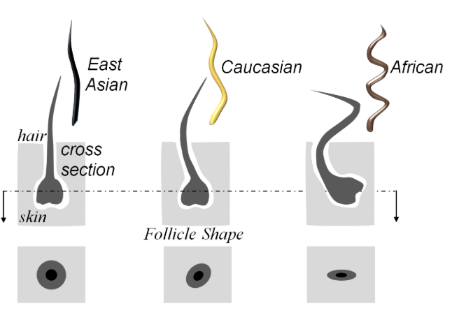
Standard EEG electrodes are unable to reliably measure electrical activity in people with coarse and curly hair. Straight and curly hair differ in the shape and size of the follicle. Straight hair has rounder and larger follicles, while curly hair has elliptical and smaller hair follicles (Figure 6.4.7). This difference makes it easier to manipulate straight hair and thus reliably record from individuals with this hair type. It also makes it more difficult to record from those with coarse and curly hair, usually those of African descent. Patients with coarse and curly hair are often asked to shave their heads or EEGs are not performed due to lack of experience of dealing with coarse and curly hair. This often leads to poor-quality data, uncomfortable patient experiences, and inability to participate in research. Even more harmful, EEGs are sometimes not used on Black patients because of these difficulties, leading to a decrease in disease detection.
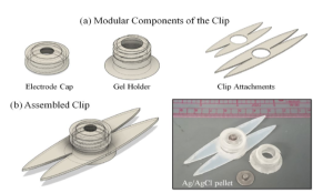
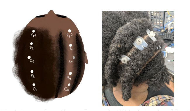
In order for neuroscience to become more inclusive, it must first examine the tools it uses to conduct research and thus advance our knowledge. Researchers at Carnegie Mellon University are working on methods to expand EEG research. Having observed the difficulties of acquiring reliable data from those with coarse and curly hair, they explored two options to improve the data. First, they studied a braiding technique that could increase the quality of the of data. The study suggests the use of thin cornrows. Cornrows are a braiding technique that exposes portions of the scalp. Second, they redesigned the shape of the electrode to better accommodate those with coarse and curly hair. Currently, an EGG is applied to the scalp by using a cap with electrodes embedded into it or small individual electrodes that are applied strategically. This current design increases noise and makes it difficult to obtain reliable data. The team designed a clip that can be attached to existing electrodes and allow for the acquirement of reliable data (Figure 6.4.8).
The combination of the cornrow braiding technique and the redesigned electrode clip design decreased electrode-skin impedance and thus decreased noise (Figure 6.4.9). There are still many ways in which research can become more inclusive; however, this research study is a step forward in the right direction.
Section Review
The adult brain is separated into four major regions: the cerebrum, the diencephalon, the brain stem, and the cerebellum. The cerebrum is the largest portion and contains the cerebral cortex and subcortical nuclei. It is divided into two halves by the longitudinal fissure.
The cortex is separated into the frontal, parietal, temporal, and occipital lobes. The frontal lobe is responsible for motor functions, from planning movements through executing commands to be sent to the spinal cord and periphery. The most anterior portion of the frontal lobe is the prefrontal cortex, which is associated with aspects of personality through its influence on motor responses in decision-making.
The other lobes are responsible for sensory functions. The parietal lobe is where somatosensation is processed. The occipital lobe is where visual processing begins, although the other parts of the brain can contribute to visual function. The temporal lobe contains the cortical area for auditory processing, but it also has regions crucial for memory formation.
Nuclei beneath the cerebral cortex, known as the subcortical nuclei, are responsible for augmenting cortical functions. The basal nuclei receive input from cortical areas and compare it with the general state of the individual through the activity of a dopamine-releasing nucleus. The output influences the activity of part of the thalamus that can then increase or decrease cortical activity that often results in changes to motor commands. The basal forebrain is responsible for modulating cortical activity in attention and memory. The limbic system includes deep cerebral nuclei that are responsible for emotion and memory.
Review Questions
Critical Thinking Questions
Glossary
- amygdala
- nucleus deep in the temporal lobe of the cerebrum that is related to memory and emotional behavior
- basal forebrain
- nuclei of the cerebrum related to modulation of sensory stimuli and attention through broad projections to the cerebral cortex, loss of which is related to Alzheimer’s disease
- basal nuclei
- nuclei of the cerebrum (with a few components in the upper brain stem and diencephalon) that are responsible for assessing cortical movement commands and comparing them with the general state of the individual through broad modulatory activity of dopamine neurons; largely related to motor functions, as evidenced through the symptoms of Parkinson’s and Huntington’s diseases
- Broca’s area
- region of the frontal lobe associated with the motor commands necessary for speech production and located only in the cerebral hemisphere responsible for language production, which is the left side in approximately 95% of the population
- caudate
- nucleus deep in the cerebrum that is part of the basal nuclei; along with the putamen, it is part of the striatum
- central sulcus
- surface landmark of the cerebral cortex that marks the boundary between the frontal and parietal lobes
- cerebral cortex
- outer gray matter covering the forebrain, marked by wrinkles and folds known as gyri and sulci
- cerebrum
- region of the adult brain that develops from the telencephalon and is responsible for higher neurological functions such as memory, emotion, and consciousness
- cerebellum
- region of the adult brain connected primarily to the pons that developed from the metencephalon (along with the pons) and is largely responsible for comparing information from the cerebrum with sensory feedback from the periphery through the spinal cord
- cerebral hemisphere
- one half of the bilaterally symmetrical cerebrum
- corpus callosum
- large white matter structure that connects the right and left cerebral hemispheres
- EEG
- A method used to measure electrical activity of the brain
- frontal eye field
- region of the frontal lobe associated with motor commands to orient the eyes toward an object of visual attention
- frontal lobe
- Region of the brain superior to the lateral sulcus and is primarily associated with motor functions and planning
- globus pallidus
- nuclei deep in the cerebrum that are part of the basal nuclei and can be divided into the internal and external segments
- hippocampus
- gray matter deep in the temporal lobe that is very important for long-term memory formation
- kinesthesia
- general sensory perception of movement of the body
- lateral sulcus
- surface landmark of the cerebral cortex that marks the boundary between the temporal lobe and the frontal and parietal lobes
- limbic cortex
- collection of structures of the cerebral cortex that are involved in emotion, memory, and behavior and are part of the larger limbic system
- limbic system
- structures at the edge (limit) of the boundary between the forebrain and hindbrain that are most associated with emotional behavior and memory formation
- longitudinal fissure
- large separation along the midline between the two cerebral hemispheres
- occipital lobe
- the posterior region of the cortex and is responsible for primary visual perception
- olfaction
- special sense responsible for smell, which has a unique, direct connection to the cerebrum
- parietal lobe
- region of the cerebral cortex associated with somatosensation
- parieto-occipital sulcus
- groove in the cerebral cortex representing the border between the parietal and occipital cortices
- postcentral gyrus
- ridge just posterior to the central sulcus, in the parietal lobe, where somatosensory processing initially takes place in the cerebrum
- precentral gyrus
- the most posterior region of the frontal lobe, also known as the primary motor cortex
- prefrontal lobe
- specific region of the frontal lobe anterior to the more specific motor function areas, which can be related to the early planning of movements and intentions to the point of being personality-type functions
- premotor area
- region of the frontal lobe responsible for planning movements that will be executed through the primary motor cortex
- proprioception
- general sensory perceptions providing information about location and movement of body parts; the “sense of the self”
- putamen
- nucleus deep in the cerebrum that is part of the basal nuclei; along with the caudate, it is part of the striatum
- somatosensation
- general senses related to the body, usually thought of as the senses of touch, which would include pain, temperature, and proprioception
- striatum
- the caudate and putamen collectively, as part of the basal nuclei, which receive input from the cerebral cortex
- subcortical nucleus
- all the nuclei beneath the cerebral cortex, including the basal nuclei and the basal forebrain
- temporal lobe
- region of the cerebral cortex associated with primary auditory sensation
Glossary Flashcards
References
Diverse Health Hub (Global, US). Making EEG Testing More Inclusive for All Hair Types. Cited: 2025 Jan 06.
Etienne, A., Laroia, T., Weigle, H., Afelin, A., Kelly, S. K., Krishnan, A., & Grover, P. (2020). Novel Electrodes for Reliable EEG Recordings on Coarse and Curly Hair. 2020 42nd Annual International Conference of the IEEE Engineering in Medicine & Biology Society (EMBC), 6151–6154.
Lim, A., “Open Neuroscience Initiative” (2021). College of Science and Health Full Text Publications. 2.
This work, Human Physiology, is adapted from Anatomy & Physiology by OpenStax, licensed under CC BY. This edition, with revised content and artwork, is licensed under CC BY-SA except where otherwise noted.
Images from Anatomy & Physiology by OpenStax are licensed under CC BY except where otherwise noted.
Access the original for free at OpenStax.
Image Descriptions
Figure 6.4.1. This simplified side-view diagram illustrates the four major regions of the human adult brain, though only three are visible from this perspective. The cerebrum (forebrain), which comprises the largest portion of the brain, occupies the upper and front areas and is divided into colored sections—beige at the front, blue on top, pink in the middle-lower area, and a small green portion at the back—representing its characteristic folded surface lobes. The cerebellum (hindbrain), shown in purple at the lower back of the brain, sits beneath the rear portion of the cerebrum and has a distinct rounded, compact structure. The brainstem, depicted in pale yellow, extends downward from the base of the brain, connecting the upper brain structures to the spinal cord. The fourth major region, the diencephalon, is a deeper internal structure located between the cerebrum and brainstem that cannot be viewed in this external side view. This diagram provides a clear overview of the brain’s major anatomical divisions and their relative positions, with the cerebrum dominating the upper region, the cerebellum positioned at the back and below, and the brainstem serving as the connecting stalk to the spinal cord. [Return to Figure 6.4.1]
Figure 6.4.2. This diagram presents two different views of the human brain. The left image shows a lateral (side) view of the brain, displaying the cerebrum with its characteristic folded surface of gyri and sulci that form the cerebral cortex. A dotted line traces the outline of the corpus callosum, a thick bundle of nerve fibers that connects the brain’s two hemispheres and lies deep within the brain’s interior. Below the cerebrum, the cerebellum is visible with its distinctive fine, parallel folds. The right image shows an anterior (front) view of the brain, revealing how the cerebrum is divided into two distinct halves: the right hemisphere on the left side of the image and the left hemisphere on the right side. Running vertically down the center between these two hemispheres is the longitudinal fissure, a deep groove that separates the left and right sides of the brain. Both views illustrate the brain in beige tones, emphasizing its folded surface texture. The spinal cord extends downward from the base of the brain in both views. These complementary perspectives demonstrate the brain’s bilateral symmetry and the complex folding patterns of the cortex that are visible from different angles. [Return to Figure 6.4.2]
Figure 6.4.3. This side-view diagram illustrates the four major lobes of the cerebral cortex and key surface landmarks. The frontal lobe, shown in pink at the front of the brain, is the largest and extends from the front of the head back to approximately the middle of the brain. Just behind it, separated by the central sulcus (a prominent groove running from top toward the side), lies the parietal lobe shown in gray at the top-rear portion. The precentral gyrus, a raised ridge of tissue immediately in front of the central sulcus, and the postcentral gyrus, directly behind it, are labeled as important functional areas for motor control and sensory processing respectively. The occipital lobe, shown in pale green at the very back of the brain, is the smallest lobe and is separated from the parietal lobe by the parieto-occipital sulcus. The temporal lobe, shown in blue-gray on the lower side of the brain, extends along the side and is separated from the frontal and parietal lobes above it by the lateral sulcus (also called the Sylvian fissure), a deep horizontal groove. Below all of these structures, the cerebellum appears in beige with its characteristic fine parallel folds, and the spinal cord extends downward. Each lobe has distinct functions: the frontal lobe handles reasoning and movement, the parietal lobe processes sensory information, the temporal lobe manages hearing and memory, and the occipital lobe processes vision. [Return to Figure 6.4.3]
Figure 6.4.4. This side-view diagram maps the functional areas of the cerebral cortex, color-coded by their roles. The frontal region contains motor areas shown in tan and olive green, including the primary motor cortex (a narrow band controlling voluntary movement), the motor association area (involved in planning movements), the frontal eye field (controlling eye movements), and Broca’s area (circled in the lower frontal region, responsible for speech production). The top-central area, shown in magenta and pink, contains sensory areas including the primary somatosensory cortex (processing touch, pain, and body position), the sensory association area (interpreting sensory information), and Wernicke’s area (involved in language comprehension). The back of the brain, shown in pink and beige, contains visual processing areas including the primary visual cortex at the very rear and the visual association area that interprets visual information. A general interpretation area is marked with a dotted circle where the parietal, temporal, and occipital lobes meet, integrating information from multiple senses. The lower side of the brain contains auditory areas shown in pink and beige, including the primary auditory cortex (processing sound) and the auditory association area (interpreting sounds and language). The cerebellum appears at the bottom in beige. This diagram demonstrates how different regions of the cortex are specialized for distinct functions, with primary areas receiving or sending basic information and association areas performing higher-level processing and integration. [Return to Figure 6.4.4]
Figure 6.4.5. This side-view diagram of the brain highlights two critical language processing areas and their connection. Broca’s area, located in the lower frontal region of the brain, is responsible for speech production and the physical formation of words. Wernicke’s area, positioned in the posterior temporal-parietal region toward the back of the brain, handles language comprehension and understanding the meaning of words. The diagram shows these two regions connected by curved lines representing the arcuate fasciculus, a bundle of nerve fibers that allows these language centers to communicate with each other. This connection enables the brain to coordinate understanding language (Wernicke’s area) with producing speech (Broca’s area), which is essential for fluent communication. The brain is shown in beige with its characteristic folded surface, and the cerebellum is visible at the bottom with its fine parallel folds. The spinal cord extends downward from the base of the brain. [Return to Figure 6.4.5]
Figure 6.4.6. This diagram shows a frontal (coronal) cross-section through the brain, as if slicing from front to back and viewing from the front. A small brain illustration at the top left indicates the plane of the section. The cross-section reveals both the outer and inner structures of the brain. The outer regions show the folded cerebral cortex with alternating beige (gray matter containing nerve cell bodies) and tan (white matter containing nerve fibers) layers creating the characteristic convoluted pattern. At the top center, the lateral ventricles appear as butterfly-shaped blue fluid-filled cavities, one in each hemisphere. Running horizontally across the middle is the corpus callosum, a thick white band of nerve fibers connecting the left and right hemispheres. Below the ventricles are deep gray matter structures collectively known as the basal ganglia, including the striatum (which comprises the caudate nucleus at the top and the putamen below it, both shown in pale blue-gray) and the globus pallidus (shown in pink), located between the putamen and the central structures. These deep nuclei play crucial roles in movement control, habit formation, and reward processing. The symmetrical nature of the brain is evident, with structures mirrored on both left and right sides. This frontal view clearly demonstrates how gray matter forms both the outer cortex and these deep nuclei, while white matter fills the space between them. [Return to Figure 6.4.6]
Figure 6.4.7. This comparative diagram illustrates how hair follicle shape differs among three major ethnic groups: East Asian, Caucasian, and African. The top row shows side-view cross-sections through the skin, depicting the hair shaft emerging from the follicle beneath the skin surface. The East Asian hair follicle is shown as nearly straight and perpendicular to the skin surface, producing straight hair. The Caucasian hair follicle has a slight curve, angled somewhat from perpendicular, producing wavy or slightly curved hair. The African hair follicle exhibits a pronounced curve or hook shape beneath the skin, angled sharply from perpendicular, which produces tightly coiled or kinky hair. The bottom row shows the corresponding cross-sectional shapes of the hair shafts themselves when cut straight across: East Asian hair appears nearly circular in cross-section, Caucasian hair is oval-shaped, and African hair is more flattened or elliptical. This structural difference in follicle shape and angle, along with the hair shaft cross-section, directly determines the natural texture and curl pattern of hair in different populations. The diagram uses simple grayscale illustrations with the skin surface clearly marked and dotted lines indicating the plane of the follicle cross-sections. [Return to Figure 6.4.7]
Figure 6.4.8. This diagram illustrates a small reusable laboratory device called a microfluidic clip, which holds tubes for fluid analysis. Part (a) shows the three modular components that make up the clip: the electrode cap (a cylindrical piece with an internal threaded opening), the gel holder (a similar cylindrical piece with threading), and the clip attachments (two elongated plastic arms or clamps). Part (b) shows the assembled clip, with the cylindrical components secured at the center where the two clip arms meet, creating a clip mechanism that can hold tubing. An accompanying photograph shows the actual device next to a coin (marked “ANG Indies”) for scale, demonstrating that the clip is quite small—only a few centimeters in length. The device appears to be made of white or light gray plastic or similar material. This modular design allows researchers to easily assemble, disassemble, and potentially replace individual components of the clip as needed for laboratory experiments involving microfluidic systems. [Return to Figure 6.4.8]
Figure 6.4.9. This two-part image compares a planned surgical approach with its real-world application. The left image shows a brown anatomical model of a human head viewed from above, with a series of white dots marking planned incision or surgical access points arranged in a curved line across the top of the scalp. The right image shows an actual patient’s head during or after a neurosurgical procedure, with the hair shaved and multiple white surgical dressings or bandages placed along the scalp following the same curved pattern as indicated in the model. The patient’s dark hair is visible around the shaved surgical area. This side-by-side comparison demonstrates how surgeons plan minimally invasive access points on models before performing procedures, and how those plans translate to actual surgical practice. The dotted pattern suggests this may be for a procedure requiring multiple small access points rather than one large incision, potentially for electrode placement, biopsy sites, or a minimally invasive surgical technique. [Return to Figure 6.4.9]
Report an Error
Did you find an error, typo, broken link, or other problem in the text? Please follow this link to the error reporting form to submit an error report to the authors.
