Chapter 6. The Central Nervous System
6.5 The Diencephalon, Brain Stem, and Cerebellum
Learning Objectives
By the end of this section, you will be able to:
- Describe the location of the diencephalon, and name its subdivisions and functions
- Identify the three regions of the brain stem and note the function of each
- Recall the functions of the reticular formation
- Describe the function of the cerebellum
In this section we will explore the diencephalon, the brain stem, and the cerebellum.
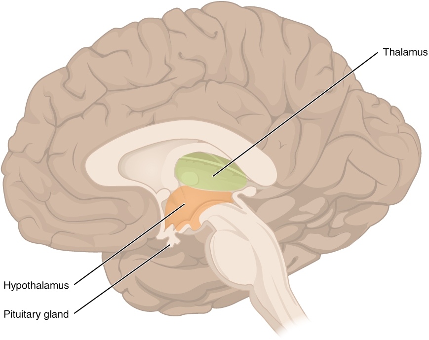
The Diencephalon
The diencephalon is the one region of the adult brain that retains its name from embryologic development. The etymology of the word diencephalon translates to “through brain.” It is the connection between the cerebrum and the rest of the nervous system, with one exception. The rest of the brain, the spinal cord, and the peripheral nervous system all send information to the cerebrum through the diencephalon. Output from the cerebrum passes through the diencephalon. The single exception is the system associated with olfaction, or the sense of smell, which connects directly with the cerebrum.
The diencephalon is deep beneath the cerebrum and constitutes the walls of the third ventricle. The diencephalon can be described as any region of the brain with “thalamus” in its name. The two major regions of the diencephalon are the thalamus itself and the hypothalamus (Figure 6.5.1). There are other structures, such as the epithalamus, which contains the pineal gland, or the subthalamus, which includes the subthalamic nucleus that is part of the basal nuclei.
Thalamus
The thalamus is a collection of nuclei that relay information between the cerebral cortex and the periphery, spinal cord, or brain stem. All sensory information, except for the sense of smell, passes through the thalamus before processing by the cortex. Axons from the peripheral sensory organs, or intermediate nuclei, synapse in the thalamus, and thalamic neurons project directly to the cerebrum. It is a requisite synapse in any sensory pathway, except for olfaction. The thalamus does not just pass the information on, it also processes that information. For example, the portion of the thalamus that receives visual information will influence what visual stimuli are important, or what receives attention.
The cerebrum also sends information down to the thalamus, which usually communicates motor commands. This involves interactions with the cerebellum and other nuclei in the brain stem. The cerebrum interacts with the basal nuclei, which involves connections with the thalamus. The primary output of the basal nuclei is to the thalamus, which relays that output to the cerebral cortex. The cortex also sends information to the thalamus that will then influence the effects of the basal nuclei.
Hypothalamus
Inferior and slightly anterior to the thalamus is the hypothalamus, the other major region of the diencephalon. The hypothalamus is a collection of nuclei that are largely involved in regulating homeostasis. The hypothalamus is the executive region in charge of the autonomic nervous system and the endocrine system through its regulation of the anterior pituitary gland. Other parts of the hypothalamus are involved in memory and emotion as part of the limbic system.
Brain Stem
The midbrain and the pons and medulla of the hindbrain are collectively referred to as the “brain stem” (Figure 6.5.2). The structure emerges from the ventral surface of the forebrain as a tapering cone that connects the brain to the spinal cord. Attached to the brain stem, but considered a separate region of the adult brain, is the cerebellum. The midbrain coordinates sensory representations of the visual, auditory, and somatosensory perceptual spaces. The pons is the main connection with the cerebellum. The pons and the medulla regulate several crucial functions, including the cardiovascular and respiratory systems.
The cranial nerves connect through the brain stem and provide the brain with the sensory input and motor output associated with the head and neck, including most of the special senses. The major ascending and descending pathways between the spinal cord and brain, specifically the cerebrum, pass through the brain stem. A diffuse region of gray matter throughout the brain stem, known as the reticular formation, is related to sleep and wakefulness, such as general brain activity and attention.
Midbrain
One of the original regions of the embryonic brain, the midbrain is a small region between the thalamus and pons. It is separated into the tectum and tegmentum, from the Latin words for roof and floor, respectively. The cerebral aqueduct passes through the center of the midbrain, such that these regions are the roof and floor of that canal.
The tectum is composed of four bumps known as the colliculi (singular colliculus), which means “little hill” in Latin. The inferior colliculus is the inferior pair of these enlargements and is part of the auditory brain stem pathway. Neurons of the inferior colliculus project to the thalamus, which then sends auditory information to the cerebrum for the conscious perception of sound. The superior colliculus is the superior pair and combines sensory information about visual space, auditory space, and somatosensory space. Activity in the superior colliculus is related to orienting the eyes to a sound or touch stimulus. If you are walking along the sidewalk on campus and you hear chirping, the superior colliculus coordinates that information with your awareness of the visual location of the tree right above you. That is the correlation of auditory and visual maps. If you suddenly feel something wet fall on your head, your superior colliculus integrates that with the auditory and visual maps and you know that the chirping bird just relieved itself on you. You want to look up to see the culprit, but do not.
The tegmentum is continuous with the gray matter of the rest of the brain stem. Throughout the midbrain, pons, and medulla, the tegmentum contains the nuclei that receive and send information through the cranial nerves, as well as regions that regulate important functions such as those of the cardiovascular and respiratory systems.
Pons
The word pons comes from the Latin word for bridge. It is visible on the anterior surface of the brain stem as the thick bundle of white matter attached to the cerebellum. The pons is the main connection between the cerebellum and the brain stem. The bridge-like white matter is only the anterior surface of the pons; the gray matter beneath that is a continuation of the tegmentum from the midbrain. Gray matter in the tegmentum region of the pons contains neurons receiving descending input from the forebrain that is sent to the cerebellum.
Medulla
The medulla (medulla oblongata) is the region known as the myelencephalon in the embryonic brain. It is responsible for many autonomic functions and contains the cardiovascular, respiratory, vomiting, and vasomotor centers. It can control blood pressure, heart rate, and ventilation.
The Cerebellum
The cerebellum, as the name suggests, is the “little brain.” It is covered in gyri and sulci like the cerebrum, and looks like a miniature version of that part of the brain (Figure 6.5.3). The cerebellum is largely responsible for comparing information from the cerebrum with sensory feedback from the periphery through the spinal cord. It does not initiate movement but contributes to coordination, precision, and timing. Lack of coordination under the influence of alcohol is due to the strong sensitivity of the cerebellum to alcohol.
Descending fibers from the cerebrum have branches that connect to neurons in the pons. Those neurons project into the cerebellum, providing a copy of motor commands sent to the spinal cord. Sensory information from the periphery, which enters through spinal or cranial nerves, is copied to a nucleus in the medulla. Fibers from this nucleus enter the cerebellum and are compared with the descending commands from the cerebrum. If the primary motor cortex of the frontal lobe sends a command down to the spinal cord to initiate walking, a copy of that instruction is sent to the cerebellum. Sensory feedback from the muscles and joints, proprioceptive information about the movements of walking, and sensations of balance are sent to the cerebellum through the inferior olive and the cerebellum compares them. If walking is not coordinated, perhaps because the ground is uneven or a strong wind is blowing, then the cerebellum sends out a corrective command to compensate for the difference between the original cortical command and the sensory feedback. The output of the cerebellum is into the midbrain, which then sends a descending input to the spinal cord to correct the messages going to skeletal muscles.
Section Review
The diencephalon includes the thalamus and the hypothalamus, along with some other structures. The thalamus is a relay between the cerebrum and the rest of the nervous system. The hypothalamus coordinates homeostatic functions through the autonomic and endocrine systems.
The brain stem is composed of the midbrain, pons, and medulla. It controls the head and neck region of the body through the cranial nerves. There are control centers in the brain stem that regulate the cardiovascular and respiratory systems.
The cerebellum is connected to the brain stem, primarily at the pons, where it receives a copy of the descending input from the cerebrum to the spinal cord. It can compare this with sensory feedback input through the medulla and send output through the midbrain that can correct motor commands for coordination.
Review Questions
Critical Thinking Questions
Glossary
- brain stem
- consists of the midbrain and the pons and medulla
- cerebellum
- region of the adult brain connected primarily to the pons that developed from the metencephalon (along with the pons) and is largely responsible for comparing information from the cerebrum with sensory feedback from the periphery through the spinal cord
- epithalamus
- region of the diencephalon containing the pineal gland
- hypothalamus
- major region of the diencephalon that is responsible for coordinating autonomic and endocrine control of homeostasis
- inferior colliculus
- half of the midbrain tectum that is part of the brain stem auditory pathway
- medulla
- brain region which contains the cardiovascular, vomiting, and vasomotor centers
- pons
- the main connection between the cerebellum and the brain stem
- reticular formation
- diffuse region of gray matter throughout the brain stem that regulates sleep, wakefulness, and states of consciousness
- superior colliculus
- half of the midbrain tectum that combines sensory information about visual space, auditory space, and somatosensory space
- tectum
- region of the midbrain, thought of as the roof of the cerebral aqueduct, which is subdivided into the inferior and superior colliculi
- tegmentum
- region of the midbrain, thought of as the floor of the cerebral aqueduct, which continues into the pons and medulla as the floor of the fourth ventricle
- thalamus
- major region of the diencephalon that is responsible for relaying information between the cerebrum and the hindbrain, spinal cord, and periphery
Glossary Flashcards
This work, Human Physiology, is adapted from Anatomy & Physiology by OpenStax, licensed under CC BY. This edition, with revised content and artwork, is licensed under CC BY-SA except where otherwise noted.
Images from Anatomy & Physiology by OpenStax are licensed under CC BY except where otherwise noted.
Access the original for free at OpenStax.
Image Descriptions
Figure 6.5.1. This side-view cross-section of the brain reveals deep internal structures that are not visible from the surface. The outer cerebral cortex is shown in beige with its characteristic folded pattern of gyri and sulci. Within the central region of the brain, three key structures are labeled and highlighted in color. The thalamus, shown in pale green, is an egg-shaped structure located in the upper-central area of the brain. It serves as a relay station, processing and directing sensory information (except smell) to appropriate areas of the cerebral cortex. Directly below the thalamus lies the hypothalamus, shown in orange, a small but critical structure that regulates vital functions including body temperature, hunger, thirst, sleep, and hormone production. Extending downward from the hypothalamus by a thin stalk is the pituitary gland, shown in gray-tan, a pea-sized endocrine gland that produces and releases hormones controlling growth, metabolism, and reproduction throughout the body. The large pale cream area in the center represents white matter tracts, and the cerebellum is visible at the lower back portion. This diagram illustrates the hypothalamic-pituitary axis, showing how these three structures work together to regulate many of the body’s essential functions through both neural and hormonal pathways. [Return to Figure 6.5.1]
Figure 6.5.2. This side-view cross-section of the brain highlights the brainstem, the stalk-like structure connecting the brain to the spinal cord. The brainstem consists of three main regions, shown in different colors from top to bottom. The midbrain, shown in light blue, is the uppermost section located just below the large central brain structures and above the cerebellum at the back. The pons, shown in pink, is the middle bulging section that appears as a rounded protrusion at the front of the brainstem. The medulla oblongata, shown in pale purple, is the lowermost section that tapers as it transitions into the spinal cord. These three structures control many vital automatic functions: the midbrain helps coordinate eye movements and processes visual and auditory information; the pons relays signals between the cerebrum and cerebellum and helps regulate breathing; and the medulla controls critical life-sustaining functions including heart rate, breathing rhythm, and blood pressure. The cerebral cortex is shown in beige with its folded surface pattern above these structures, and the large cream-colored area represents white matter tracts. The cerebellum is visible at the lower back portion of the brain. [Return to Figure 6.5.2]
Figure 6.5.3. This two-part image illustrates the cerebellum’s anatomy and neural connections. The top illustration is a detailed line drawing showing the cerebellum from behind, situated at the posterior (back) surface of the brainstem. The cerebellum displays its characteristic tightly folded surface with parallel ridges. A cross-section reveals the deep cerebellar white matter, also called the arbor vitae (meaning “tree of life”), which has a distinctive branching, tree-like appearance radiating from the center outward to the folded gray matter surface. The pons, a bulging structure on the brainstem, is shown on the left with visible fiber bundles—this is where descending input from the cerebrum enters the cerebellum through large white matter tracts. Below the pons, the inferior olive is labeled, showing where ascending sensory input from the periphery and spinal cord enters the cerebellum. The bottom image is an MRI scan showing a side view of an actual human brain, with the cerebellum highlighted in purple at the lower back portion. This scan demonstrates the cerebellum’s position relative to the rest of the brain and brainstem. The cerebellum coordinates movement, balance, and motor learning by receiving these various inputs, processing them, and sending output signals through the midbrain that descend to the spinal cord to fine-tune motor commands. [Return to Figure 6.5.3]
Report an Error
Did you find an error, typo, broken link, or other problem in the text? Please follow this link to the error reporting form to submit an error report to the authors.
