Chapter 8. Muscle Tissue
8.2 Skeletal Muscle
Learning Objectives
By the end of this section, you will be able to:
- Name and describe the various tissues that comprise the overall structure of skeletal muscle
- Compare and contrast a muscle fascicle, a skeletal muscle fiber (myocyte, muscle cell), a myofibril, a sarcomere, and a myofilament
- Describe and diagram the arrangement and composition of myofilaments in a sarcomere, including the A-band, I-band, H-zone, Z-disc (Z-line), and M-line
- Explain the organization, structures, and functions of the following components within a skeletal muscle fiber (cell): sarcolemma, sarcoplasm, sarcoplasmic reticulum, actin, myosin, titin, nebulin, troponin, and tropomyosin
- Describe the structure and composition of a thick filament as well as a thin filament within striated muscle
- Describe the sliding filament model of muscle contraction
Each skeletal muscle is an organ that consists of various integrated tissues. These tissues include skeletal muscle fibers, blood vessels, nerve fibers, and connective tissue. Each skeletal muscle has three layers of connective tissue (called mysia) that enclose it, provide structure to the muscle, and compartmentalize the muscle fibers within the muscle (Figure 8.2.1). Each muscle is wrapped in a sheath of dense, irregular connective tissue called the epimysium, which allows a muscle to contract and move powerfully while maintaining its structural integrity. The epimysium also separates muscle from other tissues and organs in the area, allowing the muscle to move independently.
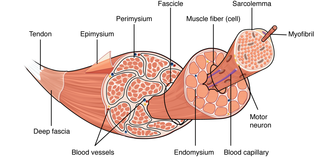
Inside each skeletal muscle, muscle fibers are organized into bundles, called fascicles, surrounded by a middle layer of connective tissue called the perimysium. This fascicular organization is common in muscles of the limbs; it allows the nervous system to trigger a specific movement of a muscle by activating a subset of muscle fibers within a fascicle of the muscle. Inside each fascicle, each muscle fiber is encased in a thin connective tissue layer of collagen and reticular fibers called the endomysium. The endomysium surrounds the extracellular matrix of the cells and plays a role in transferring force produced by the muscle fibers to the tendons.
In skeletal muscles that work with tendons to pull on bones, the collagen in the three connective tissue layers intertwines with the collagen of a tendon. At the other end of the tendon, it fuses with the periosteum coating the bone. The tension created by contraction of the muscle fibers is then transferred though the connective tissue layers, to the tendon, and then to the periosteum to pull on the bone for movement of the skeleton. In other places, the mysia may fuse with a broad, tendon-like sheet called an aponeurosis, or to fascia, the connective tissue between skin and bones. The broad sheet of connective tissue in the lower back that the latissimus dorsi muscles (the “lats”) fuse into is an example of an aponeurosis.
Every skeletal muscle is also richly supplied by blood vessels for nourishment, oxygen delivery, and waste removal. In addition, every muscle fiber in a skeletal muscle is supplied by the axon branch of a somatic motor neuron, which signals the fiber to contract. Unlike cardiac and smooth muscle, the only way to functionally contract a skeletal muscle is through signaling from the nervous system.
Skeletal Muscle Fibers
Because skeletal muscle cells are long and cylindrical, they are commonly referred to as muscle fibers (or myofibers). Skeletal muscle fibers can be quite large compared to other cells, with diameters up to 100 micrometers (μm) and lengths up to 30 centimeters (11.8 inches), as in the sartorius muscle of the upper leg. Having many nuclei allows for production of the large amounts of proteins and enzymes needed for maintaining normal function of these large protein dense cells. In addition to nuclei, skeletal muscle fibers also contain cellular organelles found in other cells, such as mitochondria and endoplasmic reticulum. However, some of these structures are specialized in muscle fibers. The specialized smooth endoplasmic reticulum, called the sarcoplasmic reticulum, stores, releases, and retrieves calcium ions (Ca++).
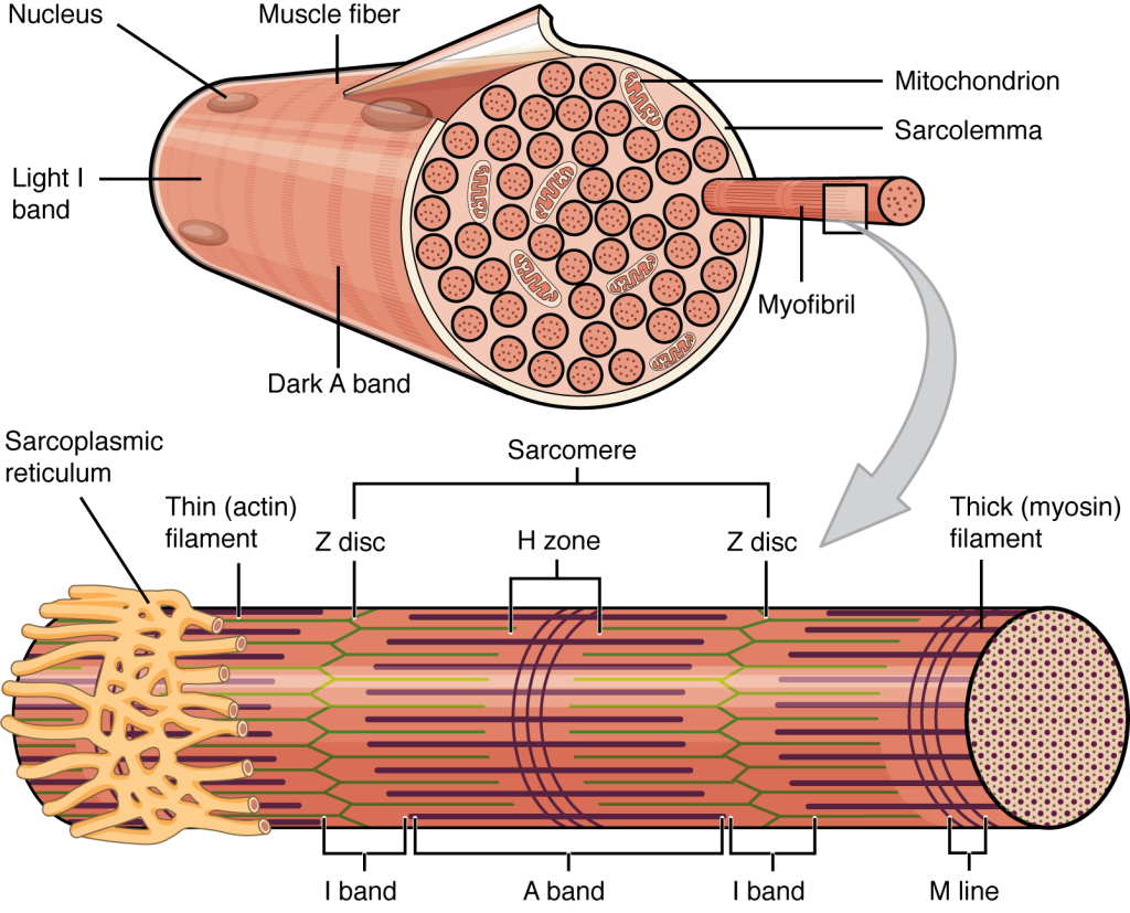
The plasma membrane of muscle fibers is called the sarcolemma (from the Greek “sarco,” which means “flesh”) and the cytoplasm is referred to as sarcoplasm (Figure 8.2.2). Within a muscle fiber, proteins are organized into organelles called myofibrils that run the length of the cell and contain sarcomeres connected in series. Because myofibrils are only approximately 1.2 μm in diameter, hundreds to thousands (each with thousands of sarcomeres) can be found inside one muscle fiber. The sarcomere, as will be described in more detail below, is the smallest functional unit of a skeletal muscle fiber and is a highly organized arrangement of the contractile myofilaments actin (thin filament) and myosin (thick filament), along with other support proteins. It is the shortening of these individual sarcomeres that leads to the contraction of individual skeletal muscle fibers (and ultimately the whole muscle).
The Sarcomere
Each sarcomere is approximately 2 μm in length with a three-dimensional cylinder-like arrangement. A sarcomere is defined as the region of a myofibril contained between two Z-discs (also called Z-lines, because pictures are two-dimensional) to which the actin myofilaments are anchored. The striated appearance of skeletal muscle fibers is due to the arrangement of the thick and thin myofilaments within each sarcomere (Figure 8.2.2). Because actin strands (projecting from the Z-discs toward the center of the sarcomere) are thinner than the myosin, they are called the thin filaments of the sarcomere. Likewise, because the myosin strands and their multiple heads have more mass and are thicker, they are called the thick filaments of the sarcomere.
The dark striated A-band is the region of the sarcomere composed of the thick (myosin) filaments, which span the center of the sarcomere and extend toward (but not all the way to) the Z-discs. Thick filaments are anchored at the middle of the sarcomere (the M-line) by a protein called myomesin. The lighter I-band regions contain thin actin filaments anchored at the Z-discs by a protein called α-actinin but do not include areas where thin and thick filaments overlap. The thin filaments extend into the A-band toward the M-line and overlap with regions of the thick filament. The A-band is dark because of the thicker myosin filaments as well as overlap with the actin filaments. The H-zone in the middle of the A-band is a little lighter in color because it only contains the portion of the thick filaments that do not overlap with the thin filaments (i.e., the thin filaments do not extend into the H-zone).
Because a sarcomere is defined by Z-discs, a single sarcomere contains one dark A-band with half of the lighter I-band on each end (Figure 8.2.2). During contraction the myofilaments themselves do not change length but actually slide across each other so the distance between the Z-discs shortens resulting in an overall shortening of the sarcomere. The length of the A-band does not change (the thick myosin filament remains a constant length), but the H-zone and I-band regions shrink. These regions represent areas where the filaments do not overlap, and as filament overlap increases during contraction, the length of these regions decrease.
Myofilament Components
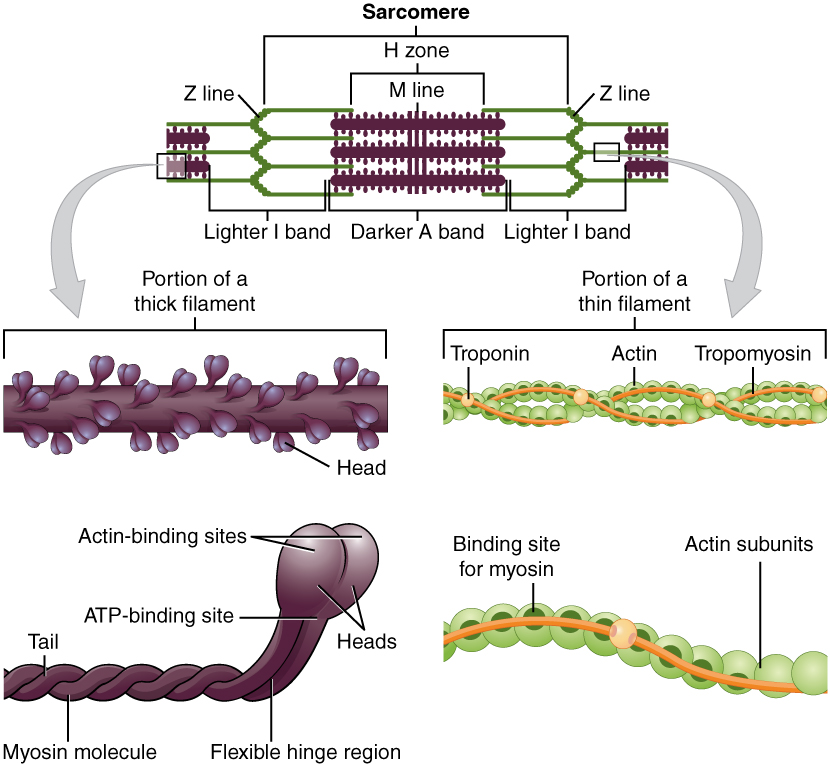
Thin filaments are composed of two filamentous actin chains (F-actin) comprised of individual actin proteins (Figure 8.2.3). These thin filaments are anchored at the Z-disc and extend toward the center of the sarcomere. Within the filament, each globular actin monomer (G-actin) contains a myosin binding site and is also associated with the regulatory proteins troponin and tropomyosin. The troponin protein complex consists of three polypeptides. Troponin I (TnI) binds to actin, troponin T (TnT) binds to tropomyosin, and troponin C (TnC) binds to calcium ions. Troponin and tropomyosin run along the actin filaments and control when the actin binding sites will be exposed for binding to myosin.
Thick filaments are composed of myosin protein complexes, which are composed of six proteins: two myosin heavy chains and four light chain molecules. The heavy chains consist of a tail region, flexible hinge region, and globular head that contains an actin-binding site and a binding site for the high energy molecule ATP. The light chains play a regulatory role at the hinge region, but the heavy chain head region interacts with actin and is the most important factor for generating force. Hundreds of myosin proteins are arranged into each thick filament with tails toward the M-line and heads extending toward the Z-discs.
Other structural proteins are associated with the sarcomere but do not play a direct role in active force production. Titin, which is the largest known protein, helps align each thick filament and adds an elastic element to the sarcomere. Titin is anchored at the M-Line, runs the length of the myosin thick filament, and extends to the Z-disc. The thin filaments also have a stabilizing protein, called nebulin, which spans the length of the thin filaments.
External Website

Watch this video to learn more about macro- and microstructures of skeletal muscles.
The Sliding Filament Model of Contraction
The arrangement and interactions between thin and thick filaments allows for the sarcomeres to generate force. When signaled by a motor neuron, a skeletal muscle fiber is activated (see Section 8.3). Cross-bridges form between the thick and thin filaments within a sarcomere, and the thin filaments are pulled past the thick filaments. This process causes the sarcomere to shorten, but the individual proteins and filaments do not change length; they simply slide along one another. This process is known as the sliding filament model of muscle contraction (Figure 8.2.4). As each sarcomere shortens, it results in a shortening of the entire skeletal muscle fiber.

The regulatory proteins troponin and tropomyosin play important roles in the sliding filament model of muscle contraction. Tropomyosin winds around the chains of the actin filament and covers myosin-binding sites on the thin filament. Tropomyosin also binds to troponin to form a troponin-tropomyosin complex, which regulates cross-bridge formation between actin and myosin microfilaments. To initiate muscle contraction, tropomyosin has to expose the myosin-binding sites on an actin filament to allow cross-bridge formation. The first step in the process is for Ca++ to bind to troponin so that tropomyosin can slide off of the binding sites on the actin strands. This allows myosin heads to bind to these exposed binding sites and form cross-bridges. The thin filaments are then pulled by the myosin heads, which slide past the thick filaments toward the center of the sarcomere. But each head can only pull a very short distance before it has reached its limit and must be “re-cocked” before it can pull again, a step that requires ATP.
Section Review
Skeletal muscles contain connective tissue, blood vessels, nerves, and muscle fibers. There are three layers of connective tissue (epimysium, perimysium, and endomysium) that help maintain the structural integrity of skeletal muscles as well as aid in its functioning. Skeletal muscle fibers are organized into groups called fascicles. Blood vessels and nerves enter the various connective tissues and branch into muscle cells.
Skeletal muscle fibers are long, multinucleated cells. The membrane of the cell is the sarcolemma; the cytoplasm of the cell is the sarcoplasm. The sarcoplasmic reticulum is a form of endoplasmic reticulum that stores, releases, and retrieves Ca++ ions. Muscle fibers are composed of myofibrils which are composed of sarcomeres linked in series. Sarcomeres are the smallest functional unit of skeletal muscle. The striations of skeletal muscle are created by the specific organization of actin and myosin filaments within sarcomeres resulting in a characteristic banding pattern. These actin and myosin filaments slide over each other to cause shortening of sarcomeres. As sarcomeres shorten, muscle fibers shorten and generate force.
Review Questions
Critical Thinking Questions
Glossary
- A-band
- the region of a sarcomere composed of the thick (myosin) filaments
- actin
- protein that makes up most of the thin myofilaments in a sarcomere muscle fiber
- aponeurosis
- broad, tendon-like sheet of connective tissue that attaches a skeletal muscle to another skeletal muscle or to a bone
- endomysium
- loose, well-hydrated connective tissue covering each muscle fiber in a skeletal muscle
- epimysium
- outer layer of connective tissue around a skeletal muscle
- fascicle
- bundle of muscle fibers within a skeletal muscle
- H-zone
- the region of a sarcomere that is in the middle of the A-band and is a little lighter in color because it only contains the portion of the thick filaments that does not overlap with the thin filaments
- I-band
- region of a sarcomere that contains thin actin filaments only; does not include areas where thin and thick filaments overlap
- M-line
- middle of a sarcomere
- myofibril
- long, cylindrical organelle that runs parallel within the muscle fiber and contains sarcomeres
- myosin
- protein that makes up most of the thick cylindrical myofilament within a sarcomere muscle fiber
- perimysium
- connective tissue that bundles skeletal muscle fibers into fascicles within a skeletal muscle
- sarcomere
- longitudinally repeating functional unit of skeletal muscle, with all of the contractile and associated proteins involved in contraction
- sarcolemma
- plasma membrane of a skeletal muscle fiber
- sarcoplasm
- cytoplasm of a muscle cell
- sarcoplasmic reticulum
- specialized smooth endoplasmic reticulum, which stores, releases, and retrieves Ca++
- thick filament
- the thick myosin strands and their multiple heads projecting from the center of the sarcomere toward (but not all to way to) the Z-discs
- thin filament
- thin strands of actin and its troponin-tropomyosin complex projecting from the Z-discs toward the center of the sarcomere
- troponin
- regulatory protein that binds to actin, tropomyosin, and calcium
- tropomyosin
- regulatory protein that covers myosin-binding sites to prevent actin from binding to myosin
Glossary Flashcards
This work, Human Physiology, is adapted from Anatomy & Physiology by OpenStax, licensed under CC BY. This edition, with revised content and artwork, is licensed under CC BY-SA except where otherwise noted.
Images from Anatomy & Physiology by OpenStax are licensed under CC BY except where otherwise noted.
Access the original for free at OpenStax.
Image Descriptions
Figure 8.2.1. This anatomical diagram illustrates the hierarchical organization of skeletal muscle tissue from macroscopic to microscopic levels. The image shows a cross-sectional view progressing from left to right, beginning with a tendon that connects to muscle tissue. The outer layer is labeled as epimysium, which surrounds the entire muscle. Within the muscle, several structures are identified: the perimysium wraps around bundles of muscle fibers called fascicles, and blood vessels are visible running through the connective tissue. The diagram shows individual muscle fibers (cells) grouped together within fascicles, each surrounded by endomysium. A magnified view on the right reveals a single muscle fiber in detail, showing the sarcolemma (cell membrane) on its surface and blood capillaries adjacent to it. A motor neuron (shown in purple) connects to the muscle fiber. The most detailed magnification displays a myofibril extending from the muscle fiber, revealing the characteristic striated pattern of repeating sarcomeres with their distinct banding. At the bottom of the image, “Deep fascia” is labeled, indicating the connective tissue layer surrounding the muscle. [Return to Figure 8.2.1]
Figure 8.2.2. This detailed diagram illustrates the hierarchical organization of skeletal muscle tissue from macroscopic to microscopic levels. The image shows a cross-sectional view progressing from left to right, beginning with a tendon that connects to muscle tissue. The outer layer is labeled as epimysium, which surrounds the entire muscle. Within the muscle, several structures are identified: the perimysium wraps around bundles of muscle fibers called fascicles, and blood vessels are visible running through the connective tissue. The diagram shows individual muscle fibers (cells) grouped together within fascicles, each surrounded by endomysium. A magnified view on the right reveals a single muscle fiber in detail, showing the sarcolemma (cell membrane) on its surface and blood capillaries adjacent to it. A motor neuron (shown in purple) connects to the muscle fiber. The most detailed magnification displays a myofibril extending from the muscle fiber, revealing the characteristic striated pattern of repeating sarcomeres with their distinct banding. At the bottom of the image, “Deep fascia” is labeled, indicating the connective tissue layer surrounding the muscle. [Return to Figure 8.2.2]
Figure 8.2.3. This multi-panel anatomical diagram illustrates the molecular structure of muscle sarcomeres and their protein components. The top panel displays a sarcomere, the basic contractile unit of muscle, with its characteristic banding pattern. The sarcomere is bounded by Z lines on either end, with a central H zone containing the M line. The lighter I bands flank the darker central A band, and the arrangement of thick filaments (shown in purple/dark) and thin filaments (shown in green) creates this striated appearance. Two curved arrows point downward from this panel to magnified views of the filament structures. The left pathway shows a “Portion of a thick filament,” depicting a myosin molecule in detail. The thick filament appears as a bundle with multiple protruding heads along its length. Below this, an individual myosin molecule is shown with its tail region, flexible hinge region, two heads with actin-binding sites, and ATP-binding site clearly labeled. The right pathway illustrates a “Portion of a thin filament,” showing the helical arrangement of actin subunits (in green) wrapped with tropomyosin (in orange) and studded with troponin complexes. A magnified view below shows the binding sites for myosin on the actin filament, with the regulatory proteins troponin and tropomyosin that control muscle contraction by exposing or blocking these binding sites. [Return to Figure 8.2.3]
Figure 8.2.4. This diagram illustrates the sliding filament mechanism of muscle contraction by comparing the molecular arrangement of sarcomeres in two states. Panel (a) shows a “relaxed sarcomere” with three parallel units displayed vertically. Each unit contains thick filaments (shown in purple with protruding myosin heads) centered between thin filaments (shown in green and yellow, representing actin with associated proteins). The sarcomere is bounded by Z lines on either end, with the central M line marking the middle of the thick filaments. The H zone, representing the region containing only thick filaments without overlap, is clearly visible in the center. Panel (b) depicts a “contracted sarcomere” where the same molecular components have moved closer together. Large black arrows pointing inward from both sides indicate the direction of contraction. In this state, the thin filaments have slid deeper between the thick filaments, causing the Z lines to move closer to the central M line and H zone. The H zone appears narrower as the actin filaments extend further into this region. The overall length of the sarcomere is noticeably shorter in the contracted state, though the lengths of the individual thick and thin filaments remain unchanged, demonstrating the sliding filament theory of muscle contraction. [Return to Figure 8.2.4]
Report an Error
Did you find an error, typo, broken link, or other problem in the text? Please follow this link to the error reporting form to submit an error report to the authors.
