Chapter 9. The Cardiovascular System: The Heart
9.5 Development of the Heart
Learning Objectives
By the end of this section, you will be able to:
- Describe the embryological development of heart structures
- Identify five regions of the fetal heart
- Relate fetal heart structures to adult counterparts
The human heart is the first functional organ to develop. It begins beating and pumping blood around day 21 or 22, a mere three weeks after fertilization. This emphasizes the critical nature of the heart in distributing blood through the vessels and the vital exchange of nutrients, oxygen, and wastes both to and from the developing baby. The critical early development of the heart is reflected by the prominent heart bulge that appears on the anterior surface of the embryo.
The heart forms from an embryonic tissue called mesoderm around 18 to 19 days after fertilization. Mesoderm is one of the three primary germ layers that differentiates early in development that collectively gives rise to all subsequent tissues and organs. The heart begins to develop near the head of the embryo in a region known as the cardiogenic area. Following chemical signals called factors from the underlying endoderm (another of the three primary germ layers), the cardiogenic area begins to form two strands called the cardiogenic cords. As the cardiogenic cords develop, a lumen rapidly develops within them. At this point, they are referred to as endocardial tubes (Figure 9.5.1). The two tubes migrate together and fuse to form a single primitive heart tube. The primitive heart tube quickly forms five distinct regions. From head to tail, these include the truncus arteriosus, bulbus cordis, primitive ventricle, primitive atrium, and the sinus venosus. Initially, all venous blood flows into the sinus venosus, and contractions propel the blood from tail to head, or from the sinus venosus to the truncus arteriosus. This is a very different pattern from that of an adult.
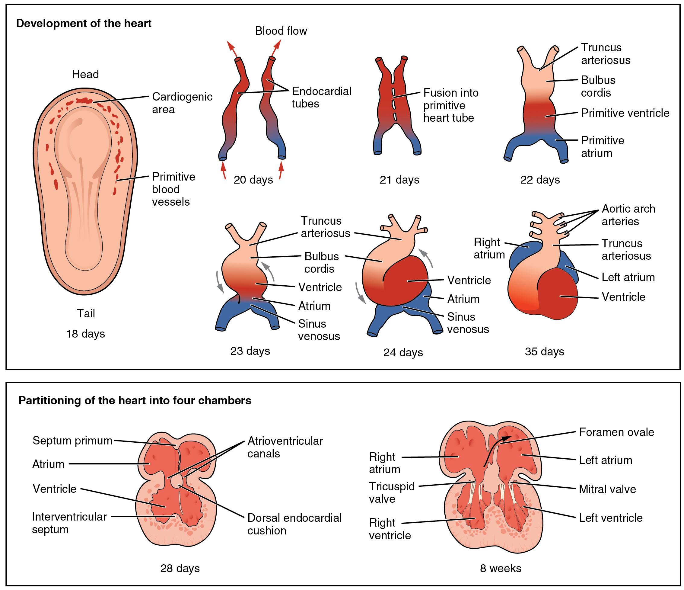
The five regions of the primitive heart tube develop into recognizable structures in a fully developed heart. The truncus arteriosus will eventually divide and give rise to the ascending aorta and pulmonary trunk. The bulbus cordis develops into the right ventricle. The primitive ventricle forms the left ventricle. The primitive atrium becomes the anterior portions of both the right and left atria, and the two auricles. The sinus venosus develops into the posterior portion of the right atrium, the SA node, and the coronary sinus.
As the primitive heart tube elongates, it begins to fold within the pericardium, eventually forming an S shape, which places the chambers and major vessels into an alignment similar to the adult heart. This process occurs between days 23 and 28. The remainder of the heart development pattern includes development of septa and valves and remodeling of the actual chambers. Partitioning of the atria and ventricles by the interatrial septum, interventricular septum, and atrioventricular septum is complete by the end of the fifth week, although the fetal blood shunts remain until birth or shortly after. The atrioventricular valves form between weeks five and eight, and the semilunar valves form between weeks five and nine.
Section Review
The heart is the first organ to form and become functional. It originates about day 18 or 19 from the mesoderm and begins beating and pumping blood about day 21 or 22. It forms from the cardiogenic region near the head and is visible as a prominent heart bulge on the surface of the embryo. Originally, it consists of a pair of strands called cardiogenic cords that quickly form a hollow lumen and are referred to as endocardial tubes. These then fuse into a single heart tube and differentiate into the truncus arteriosus, bulbus cordis, primitive ventricle, primitive atrium, and sinus venosus, starting about day 22. The primitive heart begins to form an S shape within the pericardium between days 23 and 28. The internal septa begin to form about day 28, separating the heart into the atria and ventricles, although the foramen ovale persists until shortly after birth. Between weeks five and eight, the atrioventricular valves form. The semilunar valves form between weeks five and nine.
Review Questions
Critical Thinking Questions
Glossary
- bulbus cordis
- portion of the primitive heart tube that will eventually develop into the right ventricle
- cardiogenic area
- area near the head of the embryo where the heart begins to develop 18 to 19 days after fertilization
- cardiogenic cords
- two strands of tissue that form within the cardiogenic area
- endocardial tubes
- stage in which lumens form within the expanding cardiogenic cords, forming hollow structures
- heart bulge
- prominent feature on the anterior surface of the embryo, reflecting early cardiac development
- mesoderm
- one of the three primary germ layers that differentiate early in embryonic development
- primitive atrium
- portion of the primitive heart tube that eventually becomes the anterior portions of both the right and left atria, and the two auricles
- primitive heart tube
- singular tubular structure that forms from the fusion of the two endocardial tubes
- primitive ventricle
- portion of the primitive heart tube that eventually forms the left ventricle
- sinus venosus
- develops into the posterior portion of the right atrium, the SA node, and the coronary sinus
- truncus arteriosus
- portion of the primitive heart that will eventually divide and give rise to the ascending aorta and pulmonary trunk
Glossary Flashcards
This work, Human Physiology, is adapted from Anatomy & Physiology by OpenStax, licensed under CC BY. This edition, with revised content and artwork, is licensed under CC BY-SA except where otherwise noted.
Images from Anatomy & Physiology by OpenStax are licensed under CC BY except where otherwise noted.
Access the original for free at OpenStax.
Image Descriptions
Figure 9.5.1. This diagram illustrates the developmental stages of the human heart from embryonic day 18 through 8 weeks, divided into two main sections. The upper section, titled “Development of the heart,” shows eight progressive stages. At 18 days, the embryo is depicted with a head and tail region, showing the cardiogenic area (red dots) and primitive blood vessels. By 20 days, paired endocardial tubes have formed, shown in red and blue with arrows indicating blood flow direction. At 21 days, these tubes fuse into a primitive heart tube. The 22-day stage shows the developing truncus arteriosus, bulbus cordis, primitive ventricle, and primitive atrium. Days 23 and 24 show further development with the ventricle, atrium, sinus venosus, bulbus cordis, and truncus arteriosus clearly differentiated. By day 35, the heart has formed distinct chambers with the right atrium, left atrium, ventricle, truncus arteriosus, and aortic arch arteries visible. The lower section, titled “Partitioning of the heart into four chambers,” shows two final developmental stages. At 28 days, the heart displays the septum primum, atrium, ventricle, interventricular septum, atrioventricular canals, and dorsal endocardial cushion. The final stage at 8 weeks shows the completed four-chambered heart with labeled structures including the foramen ovale, right atrium, left atrium, tricuspid valve, mitral valve, right ventricle, and left ventricle. Throughout the diagrams, arterial structures are shown in red or pink tones, while venous structures appear in blue, consistent with standard anatomical color coding for oxygenated and deoxygenated blood. [Return to Figure 9.5.1]
Report an Error
Did you find an error, typo, broken link, or other problem in the text? Please follow this link to the error reporting form to submit an error report to the authors.
